

A surgical case of infected cardiac myxoma
- PMID: 37228570
- PMCID: PMC10204046
- DOI: 10.1177/2050313X221144514
A surgical case of infected cardiac myxoma
Abstract
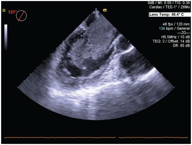
A 60-year-old woman presented with a fever of unknown origin. Echocardiography revealed a large left atrial tumor protruding into the left ventricle during diastole. Laboratory investigation showed an elevated white blood cell count, C-reactive protein concentration, and interleukin-6 concentration. Magnetic resonance imaging showed hyperacute microinfarcts and multiple old lacunar infarcts. Surgery was performed under suspicion of cardiac myxoma. A dark red jelly-like tumor with an irregular surface was removed. Histopathological examination revealed cardiac myxoma, the surface of which was covered with fibrin and bacterial masses. Preoperative blood culture was positive for Streptococcus vestibularis. These findings were compatible with a diagnosis of infected cardiac myxoma. We used an antibiotic therapeutic regimen for infective endocarditis, and the patient was discharged home on postoperative day 31. Prompt diagnosis and treatment, including effective and efficient antibiotic therapy and complete tumor resection, increased the chance of a better outcome in patients with infected cardiac myxoma.
Keywords: Cardiovascular; cardiac disease; infected cardiac myxoma; surgery.
© The Author(s) 2023.
Conflict of interest statement
The author(s) declared no potential conflicts of interest with respect to the research, authorship, and/or publication of this article.
Figures


Similar articles
-
Infected left atrial myxoma with bacteremia simulating infective endocarditis.Arch Intern Med. 1979 Oct;139(10):1176-8. Arch Intern Med. 1979. PMID: 485751
-
[Surgical treatment of infected left atrial myxoma--a case report].Nihon Kyobu Geka Gakkai Zasshi. 1992 Dec;40(12):2236-40. Nihon Kyobu Geka Gakkai Zasshi. 1992. PMID: 1491206 Review. Japanese.
-
Giant Left Atrial Myxoma with Mitral Valve Obstruction.J Cardiovasc Echogr. 2021 Apr-Jun;31(2):110-112. doi: 10.4103/jcecho.jcecho_111_20. Epub 2021 Jul 28. J Cardiovasc Echogr. 2021. PMID: 34485040 Free PMC article.
-
A Case of Infected Left Atrial Myxoma Presenting as ST-Elevation Myocardial Infarction (STEMI).Am J Case Rep. 2019 Dec 24;20:1930-1935. doi: 10.12659/AJCR.918192. Am J Case Rep. 2019. PMID: 31871313 Free PMC article.
-
Case report: left atrial Myxoma causing elevated C-reactive protein, fatigue and fever, with literature review.BMC Cardiovasc Disord. 2020 Mar 5;20(1):119. doi: 10.1186/s12872-020-01397-1. BMC Cardiovasc Disord. 2020. PMID: 32138674 Free PMC article. Review.
References
Publication types
LinkOut - more resources
Full Text Sources
Research Materials
