. 2023 Apr 1;21(2):e3052.
doi: 10.30498/ijb.2023.285965.3052. eCollection 2023 Apr.

Inhibition of NO Production in LPS-Stimulated Primary Rat Glial Cells by Gnidilatimonoein and Extract of Daphne mucronata

Affiliations

Inhibition of NO Production in LPS-Stimulated Primary Rat Glial Cells by Gnidilatimonoein and Extract of Daphne mucronata

Elham Bahrami Salehloo et al. Iran J Biotechnol. .

Abstract

Background: In the CNS, glial cells are involved in neuroinflammation and neuropathic pain. The glial cells are activated by a variety of pathological conditions and release pro-inflammatory mediators, including nitric oxide (NO). Overexpression of iNOS (inducible nitric oxide synthase) and extra NO is detrimental to neurophysiology and neuronal viability.

Objectives: This study aimed to examine the effect of Gnidilatimonein isolated from D. mucronata and its leaves extract (as natural phytochemicals) on NO production in the LPS-induced primary glial cells.

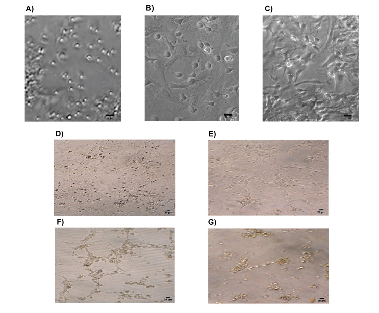
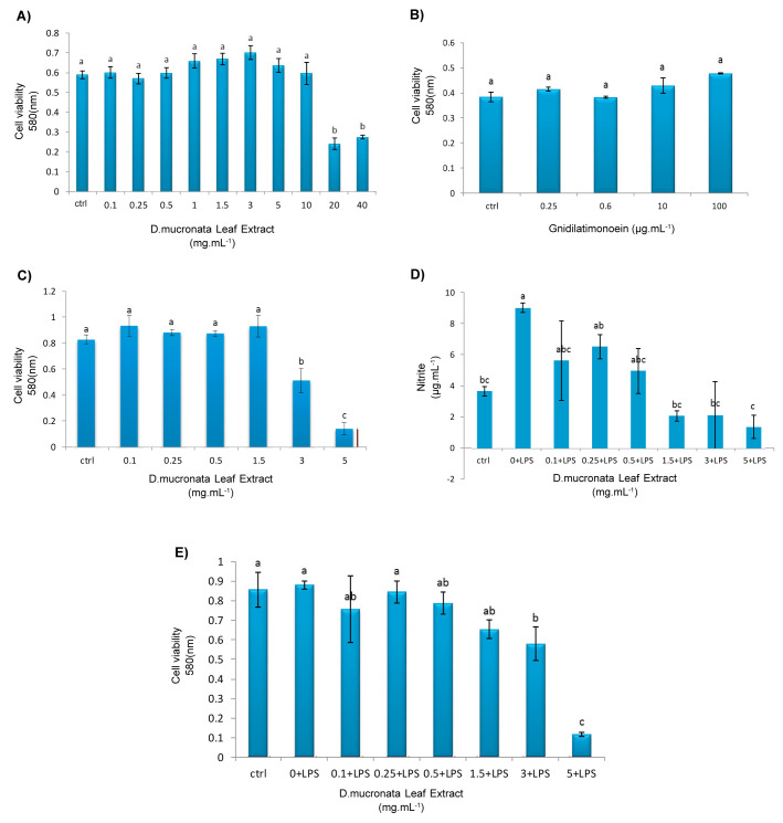
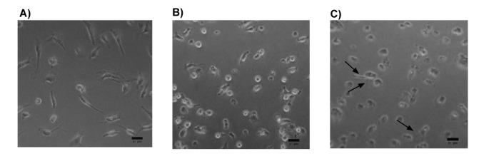
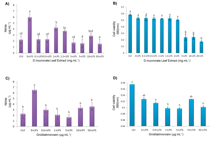
Materials and methods: A preparative HPLC method was used to isolate gnidilatimonoein from leaves ethanolic extract. Various doses of Gnidilatimonoein, the ethanolic extract were applied to primary glial cells inflamed by lipopolysaccharide. A Colorimetric test, an MTT assay, and a RT-PCR analysis were then performed to analyze and compare NO production, cell viability, and iNOS expression.

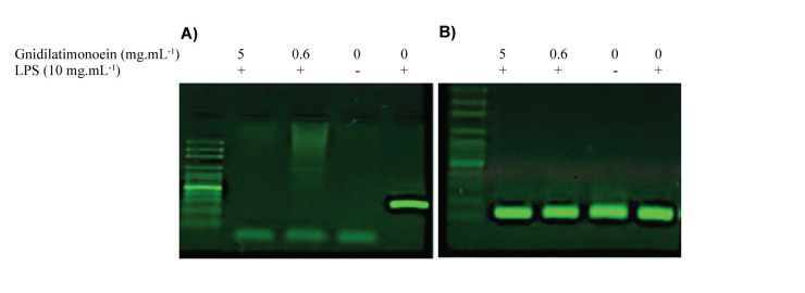
Results: Gnidilatimonoein treatment of pretreated primary glial cells significantly inhibited iNOS expression and decreased NO synthesis. Plant extracts also reduced NO production in inflamed microglial and glial at 0.1-3 mg.mL-1. At these concentrations, none of these compounds exerted a cytotoxic effect, suggesting that their anti-inflammatory effects were not due to the death of cells.

Conclusion: This study indicates that D. mucronata and its active compound, Gnidilatimonoein, could have restrained effects on the expression of iNOS on the induced glial cells; however, further investigation is warranted.

Keywords: Anti-neuroinflammation; Glial; Gnidilatimonoein; Nitric Oxide; Daphne mucronata.


Figures

Figure 1
Figure 1
Gnidilatimonoein and its Chromatogram. A) chemical structure of Gnidilatimonoein ( 20 , 23 ). B) Chromatogram of Gnidilatimonoein obtained from HPLC. (Flow gradient of Deionized water and acetonitrile, flow rate: 1 mL.min-1, the UV-Visible detector:326 nm). C) Chromatogram of Daphne mucronata leaf extract. The flash shows the peak of Gnidilatimonoein with a retention time of 16.9 min.
Figure 2
Figure 2
Morphology of Glial Cells in Different Culture Days. A) Mixed Glial cells in first, B) seventh, and C) eleventh days of culture (20x), respectively. Morphology of mixed glial cells treated with different concentrations of Daphne mucronata extract. D) Control, with x10 magnification. E) Treatment with a concentration of 5 mg.mL-1, with x20 magnification. F) treatment with 10 mg.mL-1, with x20 magnification and G) treatment with 20 mg.mL-1, with x20 magnification.
Figure 3
Figure 3
Cell viability measurement of plant extract and Gnidilatimonoein on the mixed glial cells. A) Effect of different concentrations of Daphne mucronata leaf extract (mg.mL-1) on the survival of mixed glial cells. B) Different concentrations of Gnidilatimonoein (mg.mL-1) affect the survival of mixed glial cells. Cell viability measurement of plant extract on the microglial cell. C) Effect of Daphne mucronata leaf extract on microglial cell survival. (Each column in the A, B, and C graphs, represents the average data ± standard error. Columns with at least one letter in common with the Duncan test are not significantly different at the 1% probability level). NO, and cell viability measurement of plant extract on LPS-stimulated microglial cells. D) Effects of D. mucronata leaf extract on NO production and cell viability in LPS-stimulated microglial. The cells were pretreated with various amounts of the extract before stimulation with LPS (1 mg.mL-1), and after 48 h, NO production was measured, and E) cell viability was evaluated. In the D and E graphs, Columns of at least one common letter by the Duncan test Indicate the level of significance P<0.05.
Figure 4
Figure 4
Morphology of LPS-inflamed Microglial treated with plant extract. A) Microglial in the control group. B) LPS-inflamed microglial. C) Inflamed microglial treated with 1.5 mg.mL-1 Daphne mucronata leaf extract. Photos were at 20x magnification.
Figure 5
Figure 5
NO measurement and cell viability assay of plant extract and Gnidilatimonoein on the LPS-stimulated mixed glial cells. A) Primary mixed glial cells were incubated in the absence or presence of LPS (10 mg.mL-1). The cells were pretreated with various amounts of D. mucronata extract before LPS was added. After 48h, the cultures were examined with a nitrite assay and B) a cell viability assay. C) Primary mixed glial cells were incubated in the absence or presence of LPS (10 mg.mL-1). The cells were pretreated with various amounts of Gnidilatimonoein before LPS was added. After 48h the cultures were examined with a nitrite assay and D) a cell viability assay. Columns of at least 1 common letter by the Duncan test Indicate the level of significance P<0.05.
Figure 6
Figure 6
Electrophoresis of RT-PCR productions. A) iNOS gene products, 253 bp. B) GAPDH gene products, 118 bp. Primary glial cultured were pretreated with 0.6 and 5 mg.mL-1 Gnidilatimonoein for one hour and stimulated with LPS (10 mg.mL-1) for a 48h incubation period. Gnidilatimonoein inhibits iNOS expression the same as the control group without LPS stimulation.

Similar articles

References

    1. Takeuchi H. Roles of glial cells in neuroinflammation and neurodegeneration. Clin Exp Neuroimmunol. 2013;4:2–16. doi: 10.1111/cen3.12059. - DOI
    1. Block ML, Hong JS. Microglial and inflammation-mediated neuro- degeneration: multiple triggers with a common mechanism. Prog Neurobiol. 2005;76(2):77–98. doi: 10.1016/j.pneurobio.2005.06.004. - DOI - PubMed
    1. Weydt P, Möller T. Neuroinflammation in the pathogenesis of amyotrophic lateral sclerosis. Neuroreport. 2005;16(6):527–531. - PubMed
    1. Garden GA, Moller T. Microglial biology in health and disease. J Neuroimmune Pharmacol. 2006;1(2):127–137. doi: 10.1007/s11481-006-9015-5. - DOI - PubMed
    1. Sriram K, O’Callaghan JP. Divergent roles for tumor necrosis factor-alpha in the brain. J Neuroimmune Pharmacol. 2007;2(2):140–153. doi: 10.1007/s11481-007-9070-6. - DOI - PubMed

LinkOut - more resources