. 2023 May 9:14:1166454.
doi: 10.3389/fphar.2023.1166454. eCollection 2023.

Identification of afatinib-associated ADH1B and potential small-molecule drugs targeting ADH1B for hepatocellular carcinoma

Affiliations

Identification of afatinib-associated ADH1B and potential small-molecule drugs targeting ADH1B for hepatocellular carcinoma

Yongxu Zhou et al. Front Pharmacol. .

Abstract

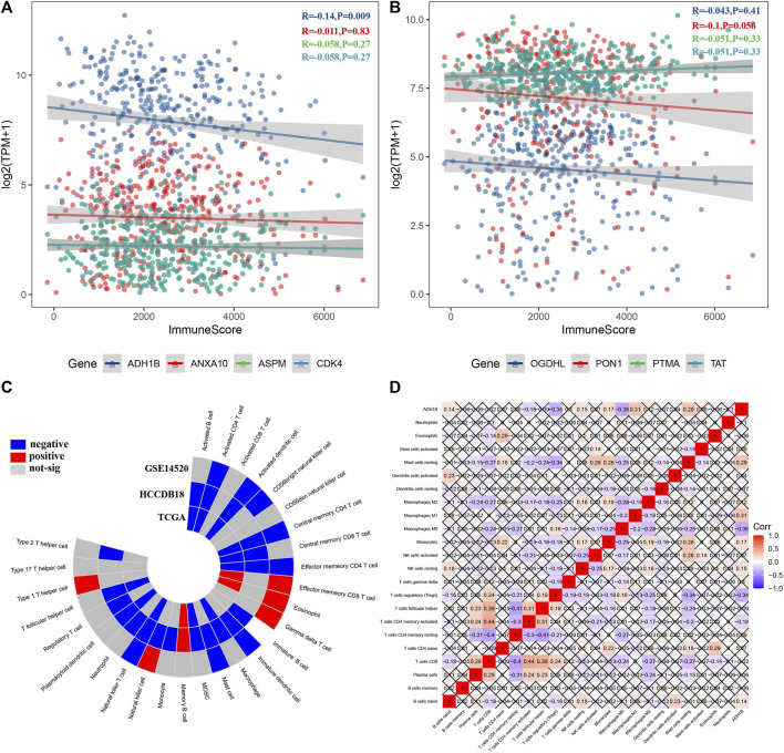
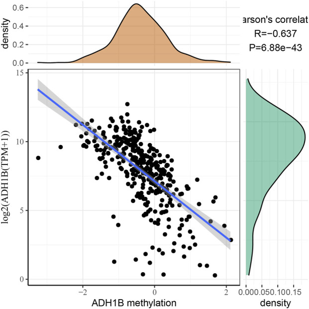
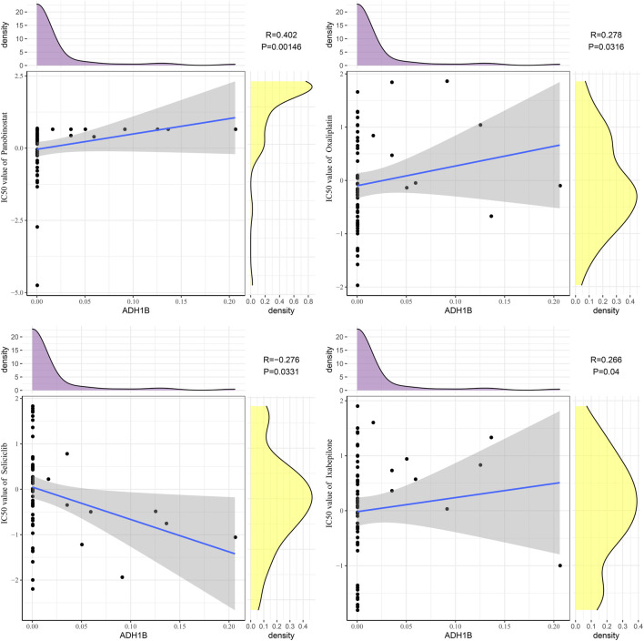
Background: Afatinib is an irreversible epidermal growth factor receptor tyrosine kinase inhibitor, and it plays a role in hepatocellular carcinoma (LIHC). This study aimed to screen a key gene associated with afatinib and identify its potential candidate drugs. Methods: We screened afatinib-associated differential expressed genes based on transcriptomic data of LIHC patients from The Cancer Genome Atlas, Gene Expression Omnibus, and the Hepatocellular Carcinoma Database (HCCDB). By using the Genomics of Drug Sensitivity in Cancer 2 database, we determined candidate genes using analysis of the correlation between differential genes and half-maximal inhibitory concentration. Survival analysis of candidate genes was performed in the TCGA dataset and validated in HCCDB18 and GSE14520 datasets. Immune characteristic analysis identified a key gene, and we found potential candidate drugs using CellMiner. We also evaluated the correlation between the expression of ADH1B and its methylation level. Furthermore, Western blot analysis was performed to validate the expression of ADH1B in normal hepatocytes LO2 and LIHC cell line HepG2. Results: We screened eight potential candidate genes (ASPM, CDK4, PTMA, TAT, ADH1B, ANXA10, OGDHL, and PON1) associated with afatinib. Patients with higher ASPM, CDK4, PTMA, and TAT exhibited poor prognosis, while those with lower ADH1B, ANXA10, OGDHL, and PON1 had unfavorable prognosis. Next, ADH1B was identified as a key gene negatively correlated with the immune score. The expression of ADH1B was distinctly downregulated in tumor tissues of pan-cancer. The expression of ADH1B was negatively correlated with ADH1B methylation. Small-molecule drugs panobinostat, oxaliplatin, ixabepilone, and seliciclib were significantly associated with ADH1B. The protein level of ADH1B was significantly downregulated in HepG2 cells compared with LO2 cells. Conclusion: Our study provides ADH1B as a key afatinib-related gene, which is associated with the immune microenvironment and can be used to predict the prognosis of LIHC. It is also a potential target of candidate drugs, sharing a promising approach to the development of novel drugs for the treatment of LIHC.

Keywords: ADH1B; CellMiner; afatinib; hepatocellular carcinoma; methylation; small-molecule drugs.


Conflict of interest statement

The authors declare that the research was conducted in the absence of any commercial or financial relationships that could be construed as a potential conflict of interest.

Figures

FIGURE 1
FIGURE 1
Screening of potential candidate genes of afatinib. (A) Volcano plots displaying 2356 differential upregulated genes and 462 downregulated differential genes between LIHC tumor and para-carcinoma tissue samples. (B, C) Venn diagram showing 178 risk genes and 42 protect genes among TCGA, HCCDB18, and GSE14520 datasets. (D) Scatter plots of correlation analysis between 220 genes and IC50 value of afatinib from LIHC cells in the GDSC2 database. (E-H) The GO and KEGG analysis of differentially expressed genes.
FIGURE 2
FIGURE 2
Distribution of eight candidate genes in clinicopathological features. The expression levels of ADH1B (A), ANXA10 (B), ASPM (C), CDK4 (D), OGDHL (E), PON1 (F), PTMA (G), and TAT (H) between early and late stage or high and low grade. ns represents p > 0.05; *p < 0.05, **p < 0.01, ***p < 0.001, and ****p < 0.0001.
FIGURE 3
FIGURE 3
Survival analysis of candidate genes. (A) Kaplan–Meier curves of eight candidate genes in the TCGA dataset. (B), Kaplan–Meier curves of eight candidate genes in the HCCDB18 dataset. (C) Kaplan–Meier curves of eight candidate genes in the GSE14520 dataset.
FIGURE 4
FIGURE 4
Identification of key genes based on immune abnormalities. (A, B) Among eight candidate genes, ADH1B was negatively correlated with the immune score. (C) ADH1B was significantly correlated with some immune cells among 28 immune cells. (D) Correlation heatmap displayed ADH1B correlated with several immune cells among 22 immune cells.
FIGURE 5
FIGURE 5
Potential regulatory pathways of ADH1B. (A) Differential enriched KEGG pathways between ADH1B-higher-expression group and ADH1B-lower-expression group. (B) GSEA showed significant enriched hallmark terms between higher ADH1B and lower ADH1B groups. (C) Scatter plots of correlation analysis between the CCP score and ADH1B. (D) Scatter plots of correlation analysis between G1/S cell cycle and ADH1B. (E) Scatter plots of correlation analysis between G2M Checkpoint and ADH1B. (F) Scatter plots of correlation analysis between inflammation pathways and ADH1B.
FIGURE 6
FIGURE 6
Correlation between ADH1B expression and ADH1B methylation. The expression of the ADH1B gene is negatively correlated with ADH1B methylation.
FIGURE 7
FIGURE 7
Drug sensitivity analysis of ADH1B. Scatter plots of correlation analysis between ADH1B and drugs (panobinostat, oxaliplatin, ixabepilone, and seliciclib).
FIGURE 8
FIGURE 8
Expression of ADH1B is decreased in the HepG2 cell line. (A) Representative Western blot results for ADH1B. (B) Quantitative analysis for ADH1B expression.

References

    1. Aldoss I. T., Tashi T., Ganti A. K. (2009). Seliciclib in malignancies. Expert Opin. investigational drugs 18 (12), 1957–1965. 10.1517/13543780903418445 - DOI - PubMed
    1. Alsfouk A. A., Alshibl H. M., Altwaijry N. A., Alsfouk B. A., Al-Abdullah E. S. (2021). Synthesis and biological evaluation of seliciclib derivatives as potent and selective CDK9 inhibitors for prostate cancer therapy. Monatsh. für Chemie-Chemical Mon. 152, 109–120. 10.1007/s00706-020-02727-x - DOI
    1. Binnewies M., Roberts E. W., Kersten K., Chan V., Fearon D. F., Merad M., et al. (2018). Understanding the tumor immune microenvironment (TIME) for effective therapy. Nat. Med. 24 (5), 541–550. 10.1038/s41591-018-0014-x - DOI - PMC - PubMed
    1. Cassetta L., Kitamura T. (2018). Macrophage targeting: Opening new possibilities for cancer immunotherapy. Immunology 155 (3), 285–293. 10.1111/imm.12976 - DOI - PMC - PubMed
    1. Charoentong P., Finotello F., Angelova M., Mayer C., Efremova M., Rieder D., et al. (2017). Pan-cancer immunogenomic analyses reveal genotype-immunophenotype relationships and predictors of response to checkpoint blockade. Cell Rep. 18 (1), 248–262. 10.1016/j.celrep.2016.12.019 - DOI - PubMed