Review
. 2023 May 25;8(1):218.
doi: 10.1038/s41392-023-01496-3.

Pathophysiological mechanisms and therapeutic approaches in obstructive sleep apnea syndrome

Affiliations
Review

Pathophysiological mechanisms and therapeutic approaches in obstructive sleep apnea syndrome

Renjun Lv et al. Signal Transduct Target Ther. .

Abstract

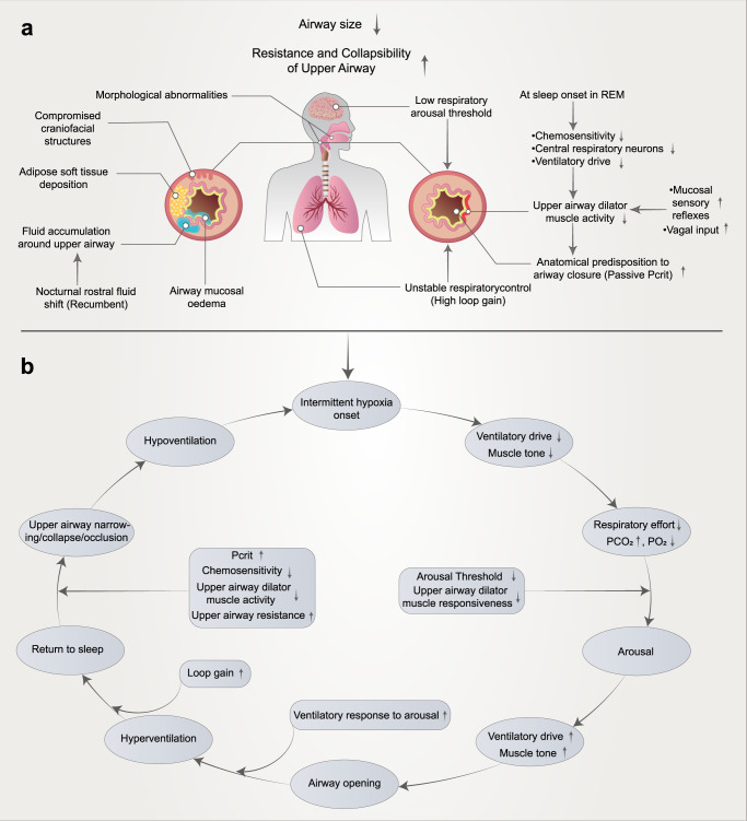
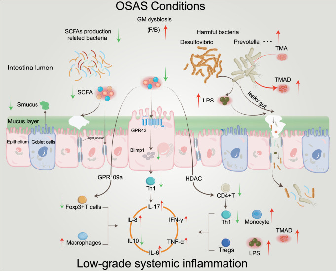
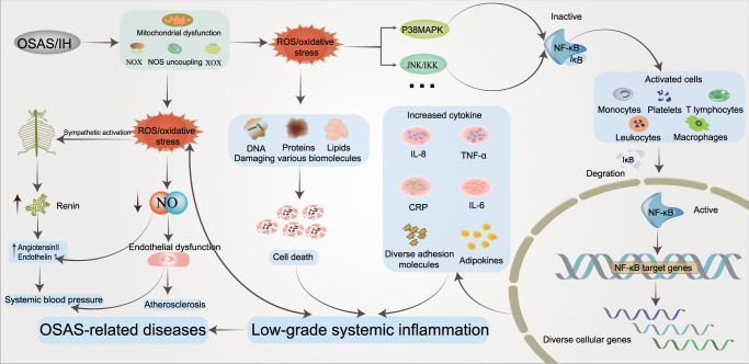
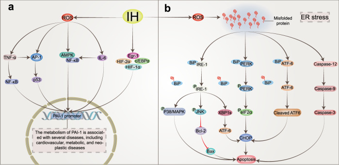
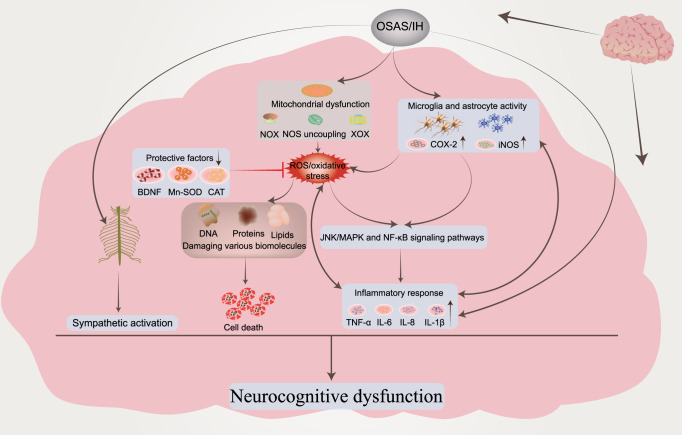
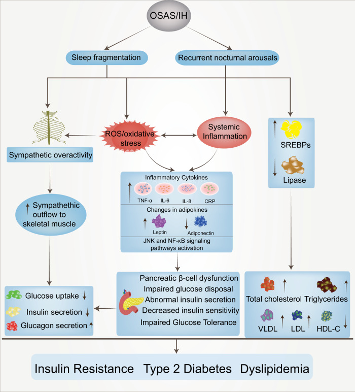
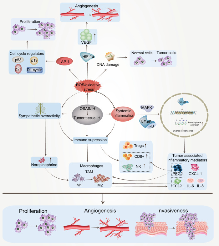
Obstructive sleep apnea syndrome (OSAS) is a common breathing disorder in sleep in which the airways narrow or collapse during sleep, causing obstructive sleep apnea. The prevalence of OSAS continues to rise worldwide, particularly in middle-aged and elderly individuals. The mechanism of upper airway collapse is incompletely understood but is associated with several factors, including obesity, craniofacial changes, altered muscle function in the upper airway, pharyngeal neuropathy, and fluid shifts to the neck. The main characteristics of OSAS are recurrent pauses in respiration, which lead to intermittent hypoxia (IH) and hypercapnia, accompanied by blood oxygen desaturation and arousal during sleep, which sharply increases the risk of several diseases. This paper first briefly describes the epidemiology, incidence, and pathophysiological mechanisms of OSAS. Next, the alterations in relevant signaling pathways induced by IH are systematically reviewed and discussed. For example, IH can induce gut microbiota (GM) dysbiosis, impair the intestinal barrier, and alter intestinal metabolites. These mechanisms ultimately lead to secondary oxidative stress, systemic inflammation, and sympathetic activation. We then summarize the effects of IH on disease pathogenesis, including cardiocerebrovascular disorders, neurological disorders, metabolic diseases, cancer, reproductive disorders, and COVID-19. Finally, different therapeutic strategies for OSAS caused by different causes are proposed. Multidisciplinary approaches and shared decision-making are necessary for the successful treatment of OSAS in the future, but more randomized controlled trials are needed for further evaluation to define what treatments are best for specific OSAS patients.


Conflict of interest statement

The authors declare no competing interests.

Figures

Fig. 1
Fig. 1
Mechanisms influencing upper airway collapse in the pathogenesis of OSAS (a) and the interplay between various factors (b). The reduction in upper airway volume caused by obesity or craniofacial structural abnormalities and soft tissue changes is an important factor in upper airway collapse. All OSAS patients have different degrees of upper airway anatomical structure injury. A nocturnal rostral fluid shift is defined as fluid accumulated in the legs during the daytime, redistributing to the upper part of the body upon lying down at night, causing an increase in peripheral pressure. In addition, most patients have mucosal edema, and the mechanism is not clear. Furthermore, several mechanisms associated with a low respiratory arousal threshold, poor pharyngeal neuromuscular muscle responsiveness, high loop gain, and high passive Pcrit may involve OSAS. When awake, neuronal activity ensures that the muscles of the dilated throat are activated, thereby preventing collapse. When this muscle loses activation during rapid eye movement (REM) sleep (chemosensitivity, central respiratory neurons, and ventilatory drive), the airway may collapse. Schematic representation of multiple pathological factors interacting to promote cyclical OSAS pathogenesis (b). In addition, these mechanisms might represent therapeutic targets. In the treatment section of this article, we introduce targeted therapies for different mechanisms
Fig. 2
Fig. 2
The mechanism of HIF‐1α activation and degradation under intermittent and continuous hypoxia conditions. Under normoxic conditions, HIF-1α is transcribed in the nucleus and translated into HIF-1α protein in the cytoplasm, which is normally hydroxylated by PHD. It then interacts with the VHL protein, undergoes ubiquitination, and is destroyed. Under continuous hypoxia, HIF-1α does not degrade but translocates to the nucleus, where it binds with HIF-1β and then recruits p300/CBP on HRE to initiate gene transcription. Among them, the HIF-1α target genes KDM4B and KDM4C were upregulated. Despite the elevated enzyme levels of KDM4A, KDM4B, and KDM4C, KDM activity was not maintained by the limited amount of oxygen, and KDM4A, KDM4B, and KDM4C remained largely inactive. This leads to increased H3K9me3, which ultimately reduces the amount of HIF-1α mRNA transcribed. Under intermittent hypoxia conditions, HIF‐1α was partially degraded during the reoxygenation phase, but the levels of KDM4B and KDM4C were increased but not to the level of continuous hypoxia. However, in contrast to continuous hypoxia, KDM4A, KDM4B, and KDM4C showed increased activity, resulting in higher H3K9me3 demethylation of the HIF‐1α gene than in normoxia or continuous hypoxia. This leads to increased production of HIF‐1α mRNA. KDMs histone lysine demethylases, H3K9me3 histone 3 lysine 9 trimethylation, HIF‐1α hypoxia-inducible factor-1, OAc acetoxy, OH hydroxyl, PHD prolyl hydroxylases, VHL von Hippel‒Lindau, EPO erythropoietin, NOS nitric oxide synthase, CBP coactivator-binding protein, HRE hypoxia response element
Fig. 3
Fig. 3
Activation of IH-associated signaling pathways. IH causes an increase in intracellular ROS, which can activate PLC-γ to produce IP3 and DAG. These two messengers are involved in intracellular signal transduction pathways and induce HIF-1α protein expression and transcriptional activity, respectively. Pathway ① indicates that IH-induced transactivation of HIF-1α requires ROS-mediated phosphorylation of the CaMKII-dependent coactivator p300. Pathway ② indicates that hypoxia-induced HIF-1α protein expression is caused by increased synthesis of mTOR, which is dependent on the ROS/Ca2+ signaling pathway. However, the mechanism by which PKC inhibits the reduction in PHD and the mechanism of the PI3K/AKT signaling pathway needs to be further confirmed (③④). Pathway ⑤ indicates that calcium-activated calpain promoted the degradation of HIF-2α protein in arterial corpuscles, resulting in a decrease in SOD2 and impaired antioxidant capacity of cells. Pathway ⑥ indicates that CaMKII can activate IEG genes, increase the transcription of c-fox mRNA or c-jun mRNA and increase the expression of AP-1, which is related to the activation of the sympathetic system and systemic inflammation. Pathway ⑦ indicates that increased ROS could stimulate the increased expression of ET and ETA and induce LTF in the carotid body. Pathway ⑧ indicates that IH causes ROS-dependent inhibition of CO production by HO-2, resulting in a decrease in PKG activity and an increase in H2S produced by CSE, which triggers a chemosensory reflex of the carotid body, leading to sympathetic excitation and hypertension. In addition, elevated H2S could activate the CAV3.2 T calcium channel on the cell membrane, causing Ca2+ influx and further aggravating the damage caused by IH (⑨). IH intermittent hypoxia, PLC-γ phospholipase C γ, PIP2 phosphatidylinositol (4,5) bisphosphate, IP3 inositol-3-phosphate, CaMKII calmodulin-dependent kinase II, IEGs immediate early genes, AP-1 activator protein-1, SOD2 superoxide dismutase 2, ET-1 endothelin 1, ETA endothelin receptor, HO-2 heme oxygenase-2, CO carbon monoxide, PKG: protein kinase G, CSE cystathionine γ-lyase, H2S hydrogen sulfide, LTF long-term facilitation
Fig. 4
Fig. 4
OSAS-induced low-grade systemic inflammation by mediating gut dysbiosis. The increased F/B ratio is a hallmark of gut microbiota dysbiosis, which is mainly characterized by a decrease in SCFA production-related bacteria and an increase in harmful bacteria. Decreased mucus secretion and mucin synthesis by dermal goblet cells disrupt the integrity of the intestinal barrier. The intestinal epithelium is dysfunctional due to inadequate nutrition, manifesting as reduced mucus production, decreased mucin secretion, and disrupted intestinal barrier integrity. Increased abundances of Prevotella and Desulfovibrio produce lipopolysaccharide and promote the degradation of mucin, increasing intestinal permeability and leading to a “leaky gut”, which triggers an intrinsic and adaptive immune response that induces low-grade inflammation in the body. Prevotella converts nutrients containing TMA into TMAO, which promotes inflammation. The reduced ability of SCFAs to activate GPR43, GPR109a, and HDAC results in diminished anti-inflammatory and increased proinflammatory capacity. GM gut microbiota, F/B Firmicutes/Bacteroidetes, SCFAs short-chain fatty acids, TMA trimethylamine, TMAO trimethylamine oxide, LPS lipopolysaccharide, HDAC histone deacetylase, GPR G-protein-coupled receptor, Blimp-1 maturation protein-1
Fig. 5
Fig. 5
Schematic demonstrating the central role played by oxidative stress and inflammation in OSAS. OSAS/IH induces ROS production by inducing mitochondrial dysfunction, activating NOX and XOX, and inducing NOS uncoupling, which results in oxidative stress. The interaction between ROS and NO further promotes oxidative stress and diminishes the bioavailability of NO, thus promoting endothelial dysfunction and inflammation, which is closely related to hypertension, atherosclerosis, and hypercoagulability. Increased ROS-dependent sympathetic activation enhances renin levels, which leads to an increase in angiotensin II, endothelin 1, and hypertension. As a second messenger, ROS can activate multiple signaling pathways (MAPK, JNK), which in turn activate NF-κB and then induce the activation of nuclear transcription factors in a variety of cells. As the main switch of the inflammatory response, NF-κB plays an important role in the pathological process of OSAS, activating and entering the nucleus, regulating the transcription of many kinds of cells (immune cells), causing an increase in cytokines and participating in the inflammatory process of cells. In addition, elevated ROS can damage intracellular macromolecular substances (DNA) and cause cell death. Various pathological processes coordinate with each other and induce low-grade inflammation in the body, which is closely related to the occurrence and progression of a variety of diseases. ROS reactive oxygen species, NOX NADPH oxidase, NOS uncoupling nitric oxide synthase uncoupling, XOX xanthine oxidase, NOS nitric oxide synthase, JNK c‐Jun N‐terminal kinase, MAPK mitogen-activated protein kinase, NF-κB nuclear factor kappa B
Fig. 6
Fig. 6
Other IH-induced signaling pathways in OSAS. IH regulates PAI-1 transcription through multiple pathways (a). IH could induce ROS, which in turn activated TNF-α, AP-1, AMPK/NF-κB pathway, and IL-6. In addition, IH could also promote the expression of Egr-1, HIF-2α, HIF-1α, and C/ΕBPα, ultimately upregulating the transcription of PAI-1. Upregulation of PAI-1 is associated with the development of IH-related disorders. Possible mechanisms of apoptosis induced by IH (b). IH could induce the generation of ROS, which in turn causes ER stress manifested by the production of misfolded proteins that bind BiP released from IRE1, PERK, and ATF6. After BiP release, IRE1, PERK, and ATF6 are activated. The activated IRE1, PERK, and ATF6 further activate their respective downstream pathways to ultimately upregulate the expression of CHOP and promote cell apoptosis. In addition, ER stress activates caspase-12, which in turn activates caspase-9 and caspase-3, leading to cell apoptosis. IH intermittent hypoxia, TNF-α tumor necrosis factor α, AP-1 activator protein-1, MAPK mitogen-activated protein kinase, NF-κB nuclear factor kappa B, C/ΕBPα CCAAT-enhancer-binding protein-α, Egr-1 early growth response protein-1, PAI-1 plasminogen activator inhibitor-1, ER endoplasmic reticulum stress, PERK protein kinase-like kinase, ATF6 transcription factor 6, IRE1 inositol requiring enzyme 1, CHOP C/EBP-homologous protein, XBP1 X-box protein-1, eIF2α e α-subunit of eukaryotic initiation factor 2
Fig. 7
Fig. 7
Proposed interactions between neurological disorders and other pathological processes induced by OSAS/IH-induced elevated ROS levels. OSAS/IH upregulates the expression of ROS in the brain, and the inhibitory effect of protective neurotrophic factors on ROS is weakened, which further leads to an increase in ROS. The macromolecular substances in injured nerve cells cause nerve cell death and activate inflammation-related signaling pathways to release inflammatory factors. Sympathetic nerve activation by OSAS/IH could cause cognitive impairment independently of other mechanisms. In addition, OSAS/IH can directly activate microglia and astrocytes and promote the release of inflammatory cytokines in the central nervous system. Excessive neuroinflammatory responses could, in turn, promote the activation of glial cells, resulting in synaptic damage and loss, neuronal necrosis, and apoptosis and ultimately leading to exaggerated neurocognitive dysfunction. BDNF brain-derived neurotrophic factor, Mn-SOD superoxide dismutase, CAT catalase, COX-2 cyclooxygenase-2, iNOS inducible nitric oxide synthase
Fig. 8
Fig. 8
Theoretical framework of possible mechanisms by which sleep fragmentation and recurrent nocturnal arousals might contribute to the occurrence of metabolic syndrome. Two key features of OSAS, namely, sleep fragmentation and recurrent nocturnal arousal, could lead to increased sympathetic nerve activity and altered glucose metabolism in skeletal muscle. ROS production in fat and activation of inflammatory pathways lead to the increased release of inflammatory factors and changes in fat-related factors, leading to metabolic dysfunction and impaired islet function. In addition, elevated SREBPs and decreased lipase caused by inflammation and oxidative stress lead to associated lipid/lipoprotein abnormalities
Fig. 9
Fig. 9
Potential mechanisms of the interaction between OSAS and cancer. IH could increase ROS levels in tumor tissues and further regulate cell cycle regulators through AP-1 to promote tumor proliferation. Elevated HIF-1α promotes the expression of VEGF and induces the growth of blood vessels in tumor tissues. The activation of NF-κB leads to the overexpression of tumor-related inflammatory mediators and tumor-related cellular immune dysfunction. In addition, enhanced sympathetic nerve activity releases norepinephrine, which can also change the tumor microenvironment and promote the occurrence of tumor cells. VEGF Vascular endothelial growth factor, CCL2 CC motif chemokine ligand 2, CXCL1 CXC motif chemokine ligand TAMs tumor-associated macrophages, M1 denotes antitumor phenotype macrophages, M2 denotes tumor-promoting phenotype macrophages

Similar articles

Cited by

References

    1. Lévy P, et al. Obstructive sleep apnoea syndrome. Nat. Rev. Dis. Prim. 2015;1:15015. doi: 10.1038/nrdp.2015.15. - DOI - PubMed
    1. Lavie L. Oxidative stress in obstructive sleep apnea and intermittent hypoxia–revisited–the bad ugly and good: implications to the heart and brain. Sleep Med. Rev. 2015;20:27–45. doi: 10.1016/j.smrv.2014.07.003. - DOI - PubMed
    1. Salzano G, et al. Obstructive sleep apnoea/hypopnoea syndrome: relationship with obesity and management in obese patients. Acta Otorhinolaryngol. Ital. 2021;41:120–130. doi: 10.14639/0392-100X-N1100. - DOI - PMC - PubMed
    1. Senaratna CV, et al. Prevalence of obstructive sleep apnea in the general population: a systematic review. Sleep Med. Rev. 2017;34:70–81. doi: 10.1016/j.smrv.2016.07.002. - DOI - PubMed
    1. Benjafield AV, et al. Estimation of the global prevalence and burden of obstructive sleep apnoea: a literature-based analysis. Lancet Respir. Med. 2019;7:687–698. doi: 10.1016/S2213-2600(19)30198-5. - DOI - PMC - PubMed

Publication types