. 2023 May 26;12(1):29.
doi: 10.1038/s41389-023-00475-1.

CDK4/6 inhibitors and the pRB-E2F1 axis suppress PVR and PD-L1 expression in triple-negative breast cancer

Affiliations

CDK4/6 inhibitors and the pRB-E2F1 axis suppress PVR and PD-L1 expression in triple-negative breast cancer

Mariusz Shrestha et al. Oncogenesis. .

Abstract

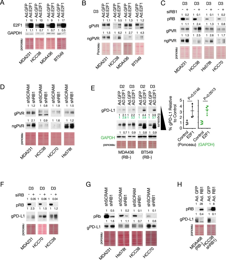
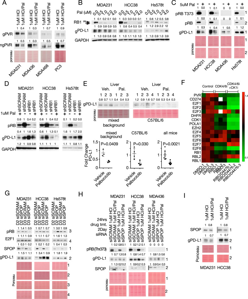
Immune-checkpoint (IC) modulators like the poliovirus receptor (PVR) and programmed death ligand 1 (PD-L1) attenuate innate and adaptive immune responses and are potential therapeutic targets for diverse malignancies, including triple-negative breast cancer (TNBC). The retinoblastoma tumor suppressor, pRB, controls cell growth through E2F1-3 transcription factors, and its inactivation drives metastatic cancer, yet its effect on IC modulators is contentious. Here, we show that RB-loss and high E2F1/E2F2 signatures correlate with expression of PVR, CD274 (PD-L1 gene) and other IC modulators and that pRB represses whereas RB depletion and E2F1 induce PVR and CD274 in TNBC cells. Accordingly, the CDK4/6 inhibitor, palbociclib, suppresses both PVR and PD-L1 expression. Palbociclib also counteracts the effect of CDK4 on SPOP, leading to its depletion, but the overall effect of palbociclib is a net reduction in PD-L1 level. Hydrochloric acid, commonly used to solubilize palbociclib, counteracts its effect and induces PD-L1 expression. Remarkably, lactic acid, a by-product of glycolysis, also induces PD-L1 as well as PVR. Our results suggest a model in which CDK4/6 regulates PD-L1 turnover by promoting its transcription via pRB-E2F1 and degradation via SPOP and that the CDK4/6-pRB-E2F pathway couples cell proliferation with the induction of multiple innate and adaptive immunomodulators, with direct implications for cancer progression, anti-CDK4/6- and IC-therapies.


Conflict of interest statement

The authors declare no competing interests.

Figures

Fig. 1
Fig. 1. RB-loss correlates with reduced expression of immune hallmarks and increased RNA expression of CD274 and PVR in mouse and human breast cancer.
A GSEA comparing immune-related pathways in RbΔp53Δ (red) with p53Δ (blue) TNBC-like mammary tumors, visualized by Cytoscape. NES normalized enrichment score. B Correlation analysis of immune markers (right labels) with molecular signatures (bottom labels) in patient samples of 1302 mixed breast cancer and 205 TNBC. C Correlation level of CD274 (PD-L1) and PVR with indicated RB-related pathways. D Summary of ENCODE analysis for the PVR and CD274 promoter region (see Supplementary Fig. S3A). *P < 0.05, **P < 0.001, ***P < 0.0001, &P = 0.0659.
Fig. 2
Fig. 2. Direct Regulation of PVR and PD-L1 by pRB and E2F1.
A Immunoblot validation of E2F1 expression following adenovirus-mediated transduction of E2F1 (Ad.E2F1) in indicated TNBC lines. B Immunoblot analysis of glycosylated and non-glycosylated PVR in multiple TNBC lines transduced with Ad.GFP or Ad.E2F1, or C depleted for RB1 via RNAi. D Immunoblot analysis of PVR response to stable depletion of RB1 by shRNA. E Left, immunoblot analysis of PD-L1 in RB1-deficient TNBC lines transduced with Ad.GFP or Ad.E2F1. Right, cumulative PD-L1 quantification normalized to ponceau (black) or to GAPDH (green). F Immunoblot analysis of PD-L1 in three different RB(+) TNBC lines following acute knockdown of RB1 via RNAi, 3 days (D3) post transfection. G Immunoblot analysis of PD-L1 in TNBC lines with stable RB1 knockdown. H Immunoblot analysis of PD-L1 in RB1-deficient (MDA-MB-468) or RB1-depleted (HCC38-shRNA) TNBC cell lines transduced with Ad.RB1. Numbers above immunoblots denote band intensity normalized to loading control.
Fig. 3
Fig. 3. The CDK4/6 inhibitor palbociclib suppresses PVR and PD-L1 expression, and counteracts the effect of SPOP depletion on PD-L1.
A Immunoblot analysis showing the effect of 2-day palbociclib treatment on ngPVR and gPVR expression. B, C Immunoblot analysis of PD-L1 in TNBC lines treated with increasing (B) or high (C) concentrations of palbociclib for 2 days. D Immunoblot analysis of PD-L1 in TNBC lines with stable knockdown of RB1, treated with/without palbociclib for 2 days. E Immunoblot analysis of PD-L1 in livers of mice on mixed background treated with palbociclib (140 mg/kg; gavage; 5 consecutive days/week) for 28 days and probed with AF1019 antibody (left) or in livers of C57BL/6 mice treated with palbociclib (140 mg/kg; gavage) for 7 consecutive days, probed with MAB90781 antibody (right). Bottom, PD-L1 quantification. Pure palbociclib (FA65120) was suspended in sodium lactate (50 nM, PH 4.0), which was used in the vehicle control mice. F Heatmap analysis of RNA-seq data (GSE177054) of MDA231 treated with ribociclib or ribociclib/D4476 for 2 days. White boxes denote out-of-range intensity. G Immunoblot analysis of PD-L1 in indicated tumor cells following knockdown of SPOP by RNAi over 2 (D2) or 3 (D3) days. H Immunoblot analysis of PD-L1 in indicated TNBC lines with/without SPOP knockdown after 2 days (D2) and treated with/without palbociclib for 24 h. I Immunoblot analysis of PD-L1 and SPOP in indicated TNBC lines after palbociclib treatment for 48 h. Numbers above immunoblots denote band intensity normalized to loading control.
Fig. 4
Fig. 4. Effect of confluency, palbociclib treatment duration, hydrochloric and lactic acids on PVR and PD-L1 expression in TNBC cells.
A Immunoblot analysis of PD-L1 in TNBC lines seeded at various densities in 6-cm plates and treated with 1 µM palbociclib for ~2 days. Bottom, PD-L1 quantification of (A) and Supplementary Fig. 7A. Statistics calculated by pairwise analysis of seeding densities and treatment durations. B Immunoblot analysis of TNBC lines treated with various concentrations of hydrochloric acid (HCl) for 2 days. Right, PD-L1 quantification and analysis by Welch’s t-test. C Immunoblot analysis of MDA231 treated with/without palbociclib and/or with/without indicated solvents. D Immunoblot analysis of PD-L1 and PVR in different TNBC lines treated with physiological concentrations of lactic acid for 2 days. Numbers above immunoblots denote band intensity normalized to loading control. E A model suggesting that in response to mitogenic signals, CDK4/6 controls PD-L1 turnover by inducing its degradation via Culin3SPOP and its transcription via pRB-E2F1. PD-L1 in red and green depicts old or newly synthesized glycosylated protein, respectively. F A model suggesting that the CDK4/6-RB-E2F axis modulates immune surveillance by transcriptionally regulating PD-L1, PVR and potentially other IC modulator genes. Solid and segmented arrows indicate validated (herein) and unvalidated targets, respectively; single arrows denote pRB and E2F1 binding sites as well as strong E2F1 recruitment by ChIP-seq analysis, whereas double arrows denote no or weak E2F1 recruitment.

References

    1. Sun C, Mezzadra R, Schumacher TN. Regulation and function of the PD-L1 checkpoint. Immunity. 2018;48:434–52. - PMC - PubMed
    1. Sanmamed MF, Chen L. A paradigm shift in cancer immunotherapy: from enhancement to normalization. Cell. 2018;175:313–26. - PMC - PubMed
    1. Johnson DB, Nebhan CA, Moslehi JJ, Balko JM. Immune-checkpoint inhibitors: long-term implications of toxicity. Nat Rev Clin Oncol. 2022;19:254–67. - PMC - PubMed
    1. Cercek A, Lumish M, Sinopoli J, Weiss J, Shia J, Lamendola-Essel M, et al. PD-1 blockade in mismatch repair-deficient, locally advanced rectal cancer. N Engl J Med. 2022;386:2363–76. - PMC - PubMed
    1. Narayan P, Wahby S, Gao JJ, Amiri-Kordestani L, Ibrahim A, Bloomquist E, et al. FDA approval summary: atezolizumab plus paclitaxel protein-bound for the treatment of patients with advanced or metastatic TNBC whose tumors express PD-L1. Clin Cancer Res. 2020;26:2284–9. - PubMed