. 2023 May 25;14(1):3027.
doi: 10.1038/s41467-023-38268-0.

Cryogenic electron ptychographic single particle analysis with wide bandwidth information transfer

Affiliations

Cryogenic electron ptychographic single particle analysis with wide bandwidth information transfer

Xudong Pei et al. Nat Commun. .

Abstract

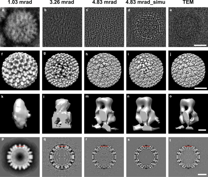
Advances in cryogenic transmission electron microscopy have revolutionised the determination of many macromolecular structures at atomic or near-atomic resolution. This method is based on conventional defocused phase contrast imaging. However, it has limitations of weaker contrast for small biological molecules embedded in vitreous ice, in comparison with cryo-ptychography, which shows increased contrast. Here we report a single-particle analysis based on the use of ptychographic reconstruction data, demonstrating that three dimensional reconstructions with a wide information transfer bandwidth can be recovered by Fourier domain synthesis. Our work suggests future applications in otherwise challenging single particle analyses, including small macromolecules and heterogeneous or flexible particles. In addition structure determination in situ within cells without the requirement for protein purification and expression may be possible.


Conflict of interest statement

The authors declare no competing interests.

Figures

Fig. 1
Fig. 1. Comparison of workflows for Cryo EPt-SPA (Left column) and TEM-SPA (Right column).
Schematic diagrams of the optical configuration a and data acquisition for ptychography b. c Typical diffraction patterns and corresponding probe positions used for ptychography (blue). Ptychographic reconstructed amplitude d and phase e of an object function. Instances of amplitude f and phase g of particles picked from d and e, respectively. h Schematic diagram of the optical configuration for TEM data acquisition. i Movie frames collected in TEM mode (left) and translations calculated for individual frames (right). j Motion-corrected image from a movie. k CTF correction by fitting the amplitude spectrum of j. l Picked particles instances from j.
Fig. 2
Fig. 2. Comparison of workflows for Cryo EPty-SPA and TEM-SPA 3D reconstructions.
ac 3D electron density maps for low, medium, and high CSAs respectively. df Corresponding bandpass filtered maps of ac, using the selected bandpass ranges where the information transfer is strongest. g Ultrawide bandwidth map obtained by Multi-band Fourier Synthesis (h) Band-limit map reconstructed by conventional TEM-SPA.
Fig. 3
Fig. 3. 3D Rotavirus DLPs reconstructions using Cryo EPty-SPA for various CSAs and conventional TEM SPA.
ac Representative experimental ptychographic phase of a virus particle with CSAs, α = 1.03, 3.26 and 4.83 mrad from sets of typically 257, 443 and 498 reconstructed particles, respectively. d Representative simulated ptychographic phase of a particle for α = 4.83 mrad from a set of typically 305 simulated particles. e Representative TEM particle image from a set of typically 378 selected particles in ref. . fj 3D maps refined with 232, 318, 241, 292 and 269 particles, respectively, corresponding to the particle instances (ae). ko Representative VP6 trimers selected from a total of 260 VP6 trimes in the outer shell of a rotavirus DLP and pt central slices (the 125th slice from 248 slices in z-direction) extracted from the 3D maps in (fj), respectively. Scale bars: 25 nm (e, j, t); 2.5 nm (o).
Fig. 4
Fig. 4. Fourier shell correlation, 3D amplitude spectrum and ultrawide bandwidth 3D map of Rotavirus DLPs.
a Fourier shell correlation curves of the 3D maps in Fig. 3f–j. b Radially averaged amplitude spectra calculated from the 3D maps in Fig. 3f–h, j and Fig. 4(c). For each spectrum for a particular CSA value a bold line segment indicates the selected bandwidth within which the information transfer is strongest at that value of the CSA. For α = 1.03 mrad, the low-frequency bandwidth from 0~0.16 nm−1 (area 1 below the bold line segment in black) is selected. For α = 3.26 and 4.83 mrad, the selected bandwidths are at medium frequency from 0.17~0.23 nm−1 (area 2 below the bold segment in red) and high frequency from 0.23~0.76 nm−1 (area 3 below the bold segment in blue), respectively. c Ultrawide bandwidth 3D map obtained by multi-band Fourier synthesis from the 3D maps in Fig. 3f–h with α = 1.03 mrad (232 particles), 3.26 mrad (318 particles) and 4.83 mrad (241 particles), respectively. Scale bars: 25 nm (c).

References

    1. Dubochet, J., Adrian, M., Chang, J.-J., Lepault, J. & McDowall, A. W. in Cryotechniques in Biological Electron Microscopy (eds R. A. Steinbrecht & K. Zierold) 114–131 (Springer Berlin Heidelberg, 1987).
    1. Frank, J. In Single-Particle Cryo-Electron Microscopy Vol. Volume 10 Series in Structural Biology 69–72 (World Scientific, 2017).
    1. Henderson R, Unwin PNT. Three-dimensional model of purple membrane obtained by electron microscopy. Nature. 1975;257:28–32. doi: 10.1038/257028a0. - DOI - PubMed
    1. Cheng Y. Membrane protein structural biology in the era of single particle cryo-EM. Curr. Opin. Struct. Biol. 2018;52:58–63. doi: 10.1016/j.sbi.2018.08.008. - DOI - PMC - PubMed
    1. Dubochet J, et al. Cryo-electron microscopy of vitrified specimens. Q. Rev. Biophys. 1988;21:129–228. doi: 10.1017/S0033583500004297. - DOI - PubMed