Review
. 2023 May 25;8(1):217.
doi: 10.1038/s41392-023-01481-w.

The blood-brain barrier: structure, regulation, and drug delivery

Affiliations
Review

The blood-brain barrier: structure, regulation, and drug delivery

Di Wu et al. Signal Transduct Target Ther. .

Abstract

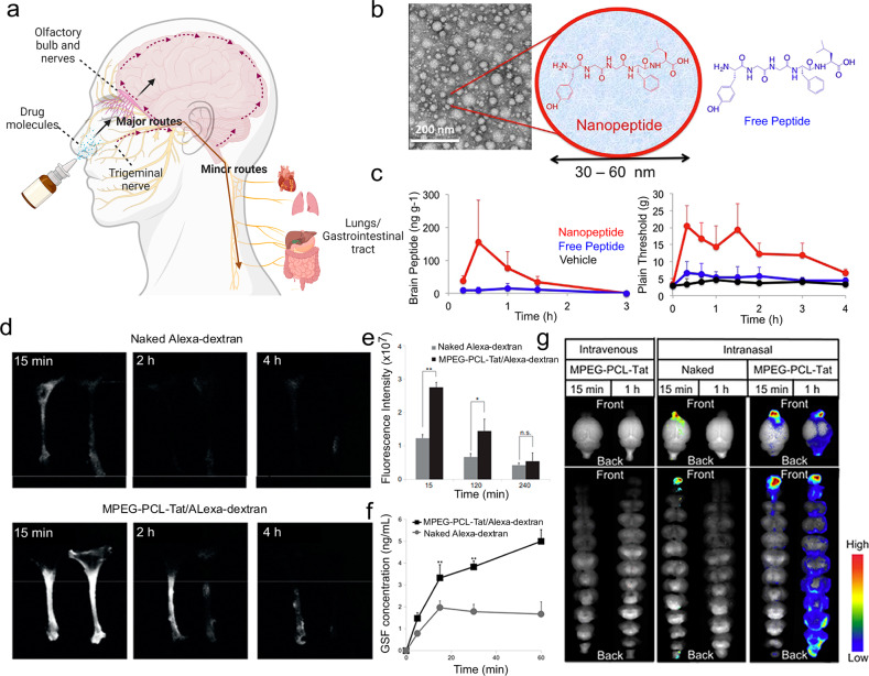
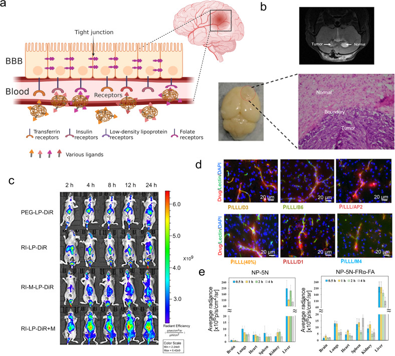
Blood-brain barrier (BBB) is a natural protective membrane that prevents central nervous system (CNS) from toxins and pathogens in blood. However, the presence of BBB complicates the pharmacotherapy for CNS disorders as the most chemical drugs and biopharmaceuticals have been impeded to enter the brain. Insufficient drug delivery into the brain leads to low therapeutic efficacy as well as aggravated side effects due to the accumulation in other organs and tissues. Recent breakthrough in materials science and nanotechnology provides a library of advanced materials with customized structure and property serving as a powerful toolkit for targeted drug delivery. In-depth research in the field of anatomical and pathological study on brain and BBB further facilitates the development of brain-targeted strategies for enhanced BBB crossing. In this review, the physiological structure and different cells contributing to this barrier are summarized. Various emerging strategies for permeability regulation and BBB crossing including passive transcytosis, intranasal administration, ligands conjugation, membrane coating, stimuli-triggered BBB disruption, and other strategies to overcome BBB obstacle are highlighted. Versatile drug delivery systems ranging from organic, inorganic, and biologics-derived materials with their synthesis procedures and unique physio-chemical properties are summarized and analyzed. This review aims to provide an up-to-date and comprehensive guideline for researchers in diverse fields, offering perspectives on further development of brain-targeted drug delivery system.


Conflict of interest statement

The authors declare no competing interests.

Figures

Fig. 1
Fig. 1
Strategies and materials for BBB regulation and brain-targeted drug delivery. a Schematic diagram of different mechanisms for BBB crossing. b Schematic diagram of BBB structure. c Engineered materials for brain-targeted drug delivery. d Various non-invasive strategies for BBB crossing
Fig. 2
Fig. 2
Intranasal administration allows rapid brain targeting from nose to brain. a Schematic illustration of the routes from the nasal cavity to the brain. b Transmission electron microscopy and structure schematic images of LENK nanoparticles. c The change of concentration of LENK in the olfactory bulb and cerebrum after the administration of nanopeptide or as the peptide alone. b, c Reproduced with permission. Copyright 2017, Elsevier. d Dynamics of fluorescence in rat trigeminal nerve. e Fluorescence intensity in the trigeminal nerve. f CSF concentration of Alexa-dextran. g Dynamics of MPEGePCLeTat complex in brain tissue. Reproduced with permission. dg Reproduced with permission. Copyright 2013, Elsevier
Fig. 3
Fig. 3
Ligands conjugation is an active targeting strategy using ligands that have high specificity toward the receptor on the endothelial cells of the brain. a Schematic illustration of receptor-mediated drug delivery using ligands conjugated nanoparticles to the brain. b The MRI images of the brain of glioma-bearing mouse. The photo and micrograph image of glioma which was removed from U87-MG glioma nude mice. c Real-time imaging of the U87-MG glioma nude mice. b, c Reproduced with permission. Copyright 2020, Ivyspring International Publisher. d Microscopic visualization of nanoconjugates after permeating the BBB. Scale bar: 20 μm. Reproduced with permission. Copyright 2022, American Chemical Society. e Biodistribution results of NIR-797-labeled NP-5N and NP-5N-FRα-FA in ICR mice. Reproduced with permission. Copyright 2019, American Chemical Society
Fig. 4
Fig. 4
Cell membranes donate drug delivery systems with the brain targeting abilities. a Schematic illustration of examples of cell membrane coating strategies. b The fluorescence images of orthotopic U87-Luc glioblastoma tumor. c In vivo pharmacokinetics. d Quantification in different organs and tumor of (doxorubicin) DOX accumulation. bd Reproduced with permission. Copyright 2018, Wiley-VCH. e The analysis of PD-1 levels on different phenotypes of macrophages. f The analysis of the PD-1+ cell ratio in different types of macrophages. g The fluorescence images pictured in groups of PLGA/DiR and PD-1-MM@PLGA/DiR. h Immunohistochemistry staining images. Scale bar: left 1 mm; right 50 μm. eh Reproduced with permission. Copyright 2022, American Chemical Society. i Schematic illustration of the process of NK@AIEdots to inhibit the growth of the brain tumor. Reproduced with permission. Copyright 2020, American Chemical Society
Fig. 5
Fig. 5
Other strategies can achieve the intention of brain-targeted drug delivery. a Schematic illustration of examples of other strategies. b Fluorescence images using in vivo imaging system of isolated brains. c Schematic illustration of the weight drop-induced TBI model. d Time points of physically invaded BBB was studied. e Schematic illustration of the isolation of primary neuronal cells from mouse embryos. be Reproduced with permission. Copyright 2020, American Association for the Advancement of Science. f Ex vivo fluorescence images of primary organs of glioma-bearing mice. g Confocal images and corresponding fluorescence intensity of 3D tumor microspheres. f, g Reproduced with permission. Copyright 2022, Nature Publishing Group. h Evaluation of general toxicity, kidney function and liver function over time. Reproduced with permission. Copyright 2022, American Association for the Advancement of Science
Fig. 6
Fig. 6
Liposomal formulations and polymeric materials for brain-targeted drug delivery. a Schematic illustration of synthesis of Apt-LP-LuH-6 liposomes and the structure characterization. Reproduced with permission. Copyright 2021, Elsevier. b Small-molecule-loaded ultrasound-controlled liposomal nanocarrier. Reproduced with permission. Copyright 2020, Nature Publishing Group. c Schematic illustration of synthesis of micelles by nanoprecipitation. Reproduced with permission. Copyright 2019, American Association for the Advancement of Science. d Schematic illustration of nanoprecipitation of peptide-modified PLGA and GALC CLEAs for the generation of enzyme delivery system. Reproduced with permission. Copyright 2021, Nature Publishing Group
Fig. 7
Fig. 7
Gold nanomaterials and carbon materials for brain-targeted drug delivery. a Simulated structures of Aβ17-36 with L- and b D-GSH-coated Au (111) surface obtained from molecular docking simulation. Reproduced with permission. Copyright 2020, Nature Publishing Group. c Schematic illustration of synthesis and d TEM images of mazindol-B6 peptide-PCB-S-curcumin-siRNA (MBPCS) and intermediate products. Reproduced with permission. Copyright 2019, Royal Society of Chemistry. e Structures and reactivity of the various nuclearities of Ru-CO clusters on GO, and f FTIR spectra of 1Ru-and 11Ru-CO. Reproduced with permission. Copyright 2018, Wiley-VCH. g Schematic illustration of synthesis of drug-loaded graphene quantum dots (GQD-D) and Cetuximab-labeled red blood cell membrane-coated GQD-D anchored inside the nanosponge (Ct-RBC@GQD-D/NS) and h their TEM images. Reproduced with permission. Copyright 2019, American Chemical Society
Fig. 8
Fig. 8
Iron oxide nanoparticles, silica nanomaterials, biomimetic nanomaterials and Cas9/RNA nanoparticles for brain-targeted drug delivery. a Schematic illustration of synthesis of Fe3O4 nanoparticles-PLGA-perfluorohexane (PFH) nanoparticles with CREKA peptides and b, c representative TEM images of Fe3O4-PLGA-PFH-CREKA and PLGA-PFH-CREKA nanoparticles, and d their elemental mapping results. Reproduced with permission. Copyright 2019, American Chemical Society. e Schematic illustration of synthesis of HA-MMSN-1F12 nanoparticles and f particle size and g TEM images of HA-MMSN and HA-MMSN-1F12 nanoparticles. Reproduced with permission. Copyright 2022, Ivyspring International Publisher. h Schematic illustration of synthesis of cell membrane-coated nanostructures and its targeted synergistic therapy of glioblastoma. Reproduced with permission. Copyright 2022, Nature Publishing Group. i Schematic illustration of synthesis of CRISPR-Cas9 nanocapsules and j their TEM images and k size distribution. Reproduced with permission. Copyright 2022, American Association for the Advancement of Science

References

    1. Abbott, N. J. Blood–brain barrier structure and function and the challenges for CNS drug delivery. J. Inherit. Metab. Dis.36, 437–449 (2013). - PubMed
    1. Abbott, N. J., Patabendige, A. A. K., Dolman, D. E. M., Yusof, S. R. & Begley, D. J. Structure and function of the blood–brain barrier. Neurobiol. Dis.37, 13–25 (2010). - PubMed
    1. Ballabh, P., Braun, A. & Nedergaard, M. The blood–brain barrier: an overview: Structure, regulation, and clinical implications. Neurobiol. Dis.16, 1–13 (2004). - PubMed
    1. Pandit, R., Chen, L. & Götz, J. The blood-brain barrier: physiology and strategies for drug delivery. Adv. Drug Deliv. Rev.165-166, 1–14 (2020). - PubMed
    1. Banks, W. A. From blood–brain barrier to blood–brain interface: new opportunities for CNS drug delivery. Nat. Rev. Drug Discov.15, 275–292 (2016). - PubMed

Publication types

Substances