. 2023 Jun;30(6):824-833.
doi: 10.1038/s41594-023-00991-z. Epub 2023 May 25.

Structural basis for pre-tRNA recognition and processing by the human tRNA splicing endonuclease complex

Affiliations

Structural basis for pre-tRNA recognition and processing by the human tRNA splicing endonuclease complex

Cassandra K Hayne et al. Nat Struct Mol Biol. 2023 Jun.

Erratum in

Abstract

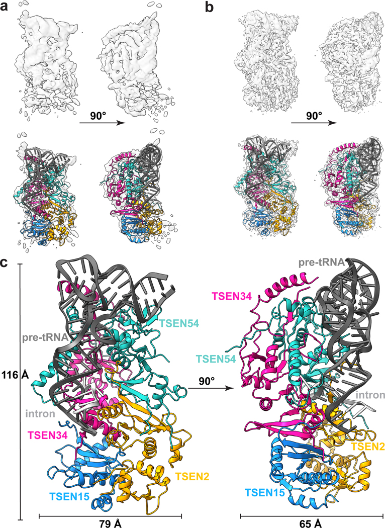
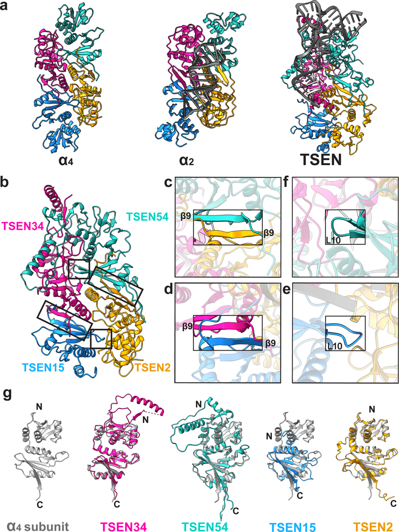
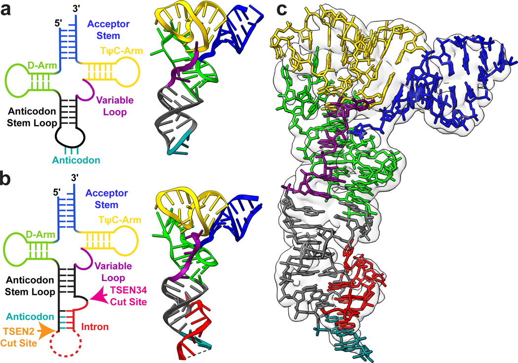
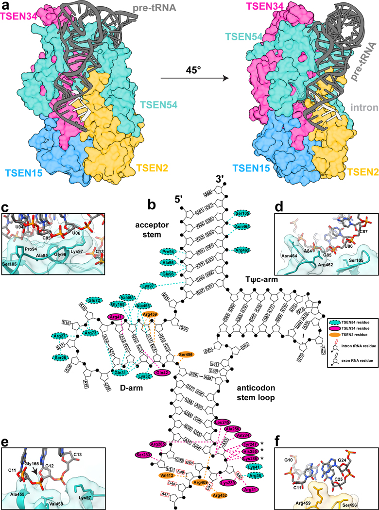
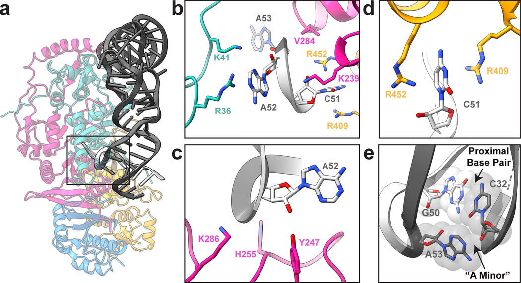
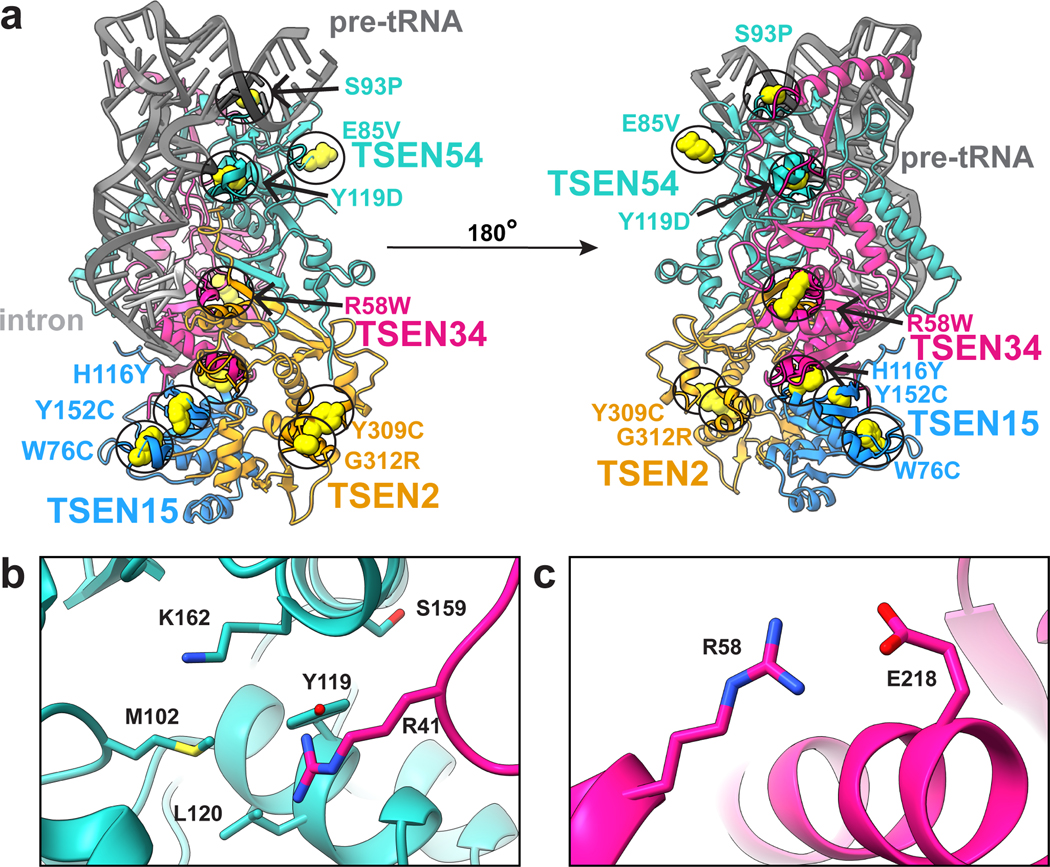
Throughout bacteria, archaea and eukarya, certain tRNA transcripts contain introns. Pre-tRNAs with introns require splicing to form the mature anticodon stem loop. In eukaryotes, tRNA splicing is initiated by the heterotetrameric tRNA splicing endonuclease (TSEN) complex. All TSEN subunits are essential, and mutations within the complex are associated with a family of neurodevelopmental disorders known as pontocerebellar hypoplasia (PCH). Here, we report cryo-electron microscopy structures of the human TSEN-pre-tRNA complex. These structures reveal the overall architecture of the complex and the extensive tRNA binding interfaces. The structures share homology with archaeal TSENs but contain additional features important for pre-tRNA recognition. The TSEN54 subunit functions as a pivotal scaffold for the pre-tRNA and the two endonuclease subunits. Finally, the TSEN structures enable visualization of the molecular environments of PCH-causing missense mutations, providing insight into the mechanism of pre-tRNA splicing and PCH.


Conflict of interest statement

Competing interests

The authors declare no conflict of interest.

Figures

Extended Data Fig. 1 |
Extended Data Fig. 1 |. WT-TSEN cryo-EM processing workflow
a. A representative micrograph, of curated 9634 micrographs. b. 1,557,097 particles were picked from the 9,634 curated micrographs, 10 representative 2D classes are shown. Particles contained in good classes were used to generate c. ab-initio reconstructions, three of which were selected and further refined using d. heterogenous refinement. The particles contained in the best three classes were further filtered using e. 2D classification and f. heterogeneous refinement prior to two classes being further refined using g. homogenous refinement, with 2x binned particles. h. A final round of heterogenous refinement resulted in one 3D class which was i. refined to an estimated 4.38 Å using homogenous refinement. j. Refinement continued with another round of homogeneous refinement resulting in a resolution of 4. Å following the unbinning of the final 161,512 particles with an estimated resolution of 4.2 Å. k. A local refinement resulted in a final map with an estimated resolution of 3.9 Å.
Extended Data Fig. 2
Extended Data Fig. 2. EndoX-TSEN structure cryo-EM processing workflow.
a. A representative micrograph of 8095 curated micrographs. b. 570,104 particles were picked from the 8059 curated micrographs and used for 2D classification. Particles from the good classes were used to generate c. ab-initio reconstructions, two of which were selected and further refined using d. heterogenous refinement. e. The particles contained in the best class were binned and refined with homogenous refinement. f. The particles were then reextracted and unbinned and used for another round of g. homogenous refinement followed by h. non-uniform refinement and i. local refinement resulting in a final map with an estimated resolution of 3.28 Å. j. FSC of the model to map
Extended Data Fig. 3
Extended Data Fig. 3. Example density for the TSEN complex.
Example densities for residues along helices from a. TSEN34, b. TSEN54, c. TSEN15, d. TSEN2, the β9-β9 interfaces for β-sheets for e. TSEN34, f. TSEN54, g. TSEN15, h. TSEN2 and density for the i. proximal base pair of the tRNA as well as the L10 loops for j. TSEN54, k. TSEN15, and the l. cation-π
Extended Data Fig. 4
Extended Data Fig. 4. BS3 crosslinking of TSEN complexes.
SDS-Page gels showing the DMSO control and crosslinked samples for a. wt-TSEN complex and b. TSEN Complex and CLP1. c-h. MSMS spectra of CLP1 peptides crosslinked to other members of the complex. Panels c (m/z 822.4292) and d (466.4795 m/z) show MSMS spectra of peptides arising from CLP1 and TSEN2. Panels e (m/z 412.2311), f (m/z 739.0369), g (m/z 671.3516), and h (m/z 398.5568) show MSMS spectra arising from crosslinks from CLP1 and TSEN54. Fragment ions that arise from green colored peptides are shown in green, fragments arising from blue colored peptides are shown in blue, and unassigned ions shown in black.
Extended Data Fig. 5
Extended Data Fig. 5. Molecular dynamics simulations account for stable complexes during dynamics.
a. Root mean square deviations (RMSD) of individual proteins and the complex averaged over each run and averaged over the five runs. TSEN34 has the largest deviations while TSEN15 shows smallest deviations irrespective of the RNA binding. Standard deviations are shown in parenthesis. The reference (Complex I) was the starting Cryo-EM configuration. Complex (II) is without tRNA and Complex (III) is without TSEN15. b. Root mean square fluctuations of individual residues averaged calculated during the microsecond dynamics and averaged over all runs. Standard deviations are shown as error bars. c. Representative dynamic cross correlation matrices of the protein components from Complex (I).
Extended Data Fig. 6
Extended Data Fig. 6. TSEN54 is the anchor that mediates the interaction of CLP1 with the TSEN complex.
Overexpression (in HEK cells) and immunoprecipitations of the individual TSEN proteins (lanes 1–4 and lanes 7–10) or full TSEN complex (lanes 5 and 11) in the absence (lanes 1–5) and presence (lanes 7–11) of CLP1 reveals strong association between CLP1 and TSEN54 (lane 10) as well as the full TSEN complex (lane 11). The experiments were conducted as previously reported using CLP1-TEV-GFP.
Extended Data Fig. 7 |
Extended Data Fig. 7 |. Structural comparison of the TSEN and EndA active site residues.
a. Overlay of TSEN + tRNA (colored as before) and EndA + BHB (grey, PDBID:2GJW) showing overall similar RNA binding. b. Overlay of the active site residues for the 3’ splice site and the c. cation-π residues from TSEN2 shown. d. Overlay of the cation-π residues from TSEN34 and the e. TSEN2 active site residues for the 5’ splice site. f. tRNA cleavage assays of a broccoli RNA -aptamer containing pre-tRNA-ILE using samples immunoprecipitated via the mutants of the 3’ splice site (pulled down by a FLAG tag on TSEN2, except for the 34 mutant) or g. 5’ splice site (pulled down by a FLAG tag on TSEN34, except for TSEN2 active site mutant). TSEN proteins that weren’t the pull-down target had a MYC-tag. The data shown is one representative experiment of three biological replicates.
Fig. 1|
Fig. 1|. Cryo-EM structures of the human TSEN complex.
Cryo-EM reconstructions of: a. wt-TSEN•pre-tRNA and b. endoX-TSEN•pre-tRNA, with cartoon overlays below. Due to its improved resolution, the endoX reconstruction was used to build and refine the overall structure. c. Model of the human TSEN complex at 0° and 90°, with overall dimensions of 116 × 79 × 65 Å. The subunits are colored as follows: TSEN54 (teal), TSEN34 (pink), TSEN2 (orange), TSEN15 (blue). The pre-tRNA is shaded in dark grey, with the intron in light grey.
Fig. 2|
Fig. 2|. The human TSEN complex retains the core architecture from the archaeal complex.
a. Structures of a homotetrameric (α4, PDBID: 1A79), homodimeric BHB bound (α2, PDBID:2GJW), and the pre-tRNA bound TSEN complex, colored to mimic the arrangement of the subunits within the TSEN complex (TSEN54 – teal, endonuclease TSEN2 - orange, endonuclease TSEN34 – pink, TSEN15 – medium blue). b. The TSEN complex retains β9-β9 interactions at the C-terminal domain of endonuclease/structural protein interfaces: c. TSEN2:TSEN54 and d. TSEN34:TSEN15. The L10-loop of the structural proteins e. TSEN54 and f. TSEN15 link the β9-β9 heterodimers together. g. A single archaeal homotetramer α subunit (PDBID: 1A79, grey) superimposed with each individual TSEN subunit.
Fig. 3 |
Fig. 3 |. Structure of the pre-tRNA reveals a familiar architecture to mature tRNA.
2D and 3D structures of a. unmodified mature tRNA-PHE (PDBID: 3L0U) and b. intron containing pre-tRNA-ARG structure, with the acceptor stem (blue), D-Arm (lime green), anticodon stem loop (dark grey), anticodon (teal), intron (red), variable loop (purple) and TψC-Arm (yellow) labeled. The cut sites for TSEN2 (orange) and TSEN34 (pink) are labelled on the pre-tRNA 2D structure. c. Electron density map around the pre-tRNA, with the pre-tRNA structure docked into the density map.
Fig. 4 |
Fig. 4 |. Recognition of pre-tRNA features by the TSEN complex.
a. Surface representation of the TSEN complex with a cartoon of the pre-tRNA, showing significant interaction between TSEN54 and multiple regions of the pre-tRNA. Colors: exons (dark grey), intron (light grey), TSEN54 (teal), TSEN34 (pink), TSEN2 (orange), TSEN15 (blue). b. Cloverleaf 2D tRNA structure with selected TSEN interfaces labeled. TSEN-tRNA interfaces mediated by hydrogen bonds and hydrophobic contacts were identified with NucPlot and visual inspection of the cryo-EM reconstruction. Regions of the tRNA from the intron are indicated with the rectangle, representing the base, boxed in red. * indicates the TSEN34 catalytic residues which were all mutated to alanine to prevent cleavage. Interfaces between TSEN54 and c. and d. the acceptor stem e. the D-arm. f. Interface between the D-arm and TSEN2.
Fig. 5 |
Fig. 5 |. Conserved catalytic core at the 3’ splice site.
a. Cartoon structure of the complex with a box highlighting the 3’ splice site. b. Overview of the three nucleotide bulge in the 3’ splice site with important interactions labeled including TSEN54 (teal) (R36 and K41), TSEN34 (pink) (V284 and K239), and TSEN2 (orange) (R409 and R452). c. Modeled positions of the catalytic residues from TSEN34 (Y247, H255, and K286), which were mutated to alanine to prevent cleavage. d. TSEN2 residues R409 and R452 form a cation-π interaction with the C51 RNA base of the intron to stabilize the bulge. e. The ASL contains the proximal base pair (C32 and G50), important for splicing in vivo. The C32 base forms an A-minor like motif with A53 from the bulge.
Fig. 6 |
Fig. 6 |. PCH mutations mapped onto the TSEN complex structure.
a. Previously reported PCH mutations are indicated with yellow spheres. b. TSEN54-Y119 is buried within the TSEN54 N-terminal domain and is in close proximity to TSEN34-R41 and several other TSEN54 (M102, L120, S159, K162) residues. c. TSEN34-R58 forms a salt bridge with TSEN34-E2.

References

    1. Hopper AK & Nostramo RT tRNA Processing and Subcellular Trafficking Proteins Multitask in Pathways for Other RNAs. Front Genet 10, 96 (2019). 10.3389/fgene.2019.00096 - DOI - PMC - PubMed
    1. Schmidt CA & Matera AG tRNA introns: Presence, processing, and purpose. Wiley Interdiscip Rev RNA 11, e1583 (2020). 10.1002/wrna.1583 - DOI - PubMed
    1. Gogakos T. et al. Characterizing Expression and Processing of Precursor and Mature Human tRNAs by Hydro-tRNAseq and PAR-CLIP. Cell Rep 20, 1463–1475 (2017). 10.1016/j.celrep.2017.07.029 - DOI - PMC - PubMed
    1. Chan PP & Lowe TM GtRNAdb 2.0: an expanded database of transfer RNA genes identified in complete and draft genomes. Nucleic Acids Res 44, D184–189 (2016). 10.1093/nar/gkv1309 - DOI - PMC - PubMed
    1. Hayne CK, Lewis TA & Stanley RE Recent insights into the structure, function, and regulation of the eukaryotic transfer RNA splicing endonuclease complex. Wiley Interdiscip Rev RNA, e1717 (2022). 10.1002/wrna.1717 - DOI - PMC - PubMed

Methods-only references:

    1. Schellenberg MJ, Petrovich RM, Malone CC & Williams RS Selectable high-yield recombinant protein production in human cells using a GFP/YFP nanobody affinity support. Protein Sci 27, 1083–1092 (2018). 10.1002/pro.3409 - DOI - PMC - PubMed
    1. Pillon MC et al. Cryo-EM reveals active site coordination within a multienzyme pre-rRNA processing complex. Nature structural & molecular biology 26, 830–839 (2019). 10.1038/s41594-019-0289-8 - DOI - PMC - PubMed
    1. Zheng SQ et al. MotionCor2: anisotropic correction of beam-induced motion for improved cryo-electron microscopy. Nat Methods 14, 331–332 (2017). 10.1038/nmeth.4193 - DOI - PMC - PubMed
    1. Punjani A, Rubinstein JL, Fleet DJ & Brubaker MA cryoSPARC: algorithms for rapid unsupervised cryo-EM structure determination. Nat Methods 14, 290–296 (2017). 10.1038/nmeth.4169 - DOI - PubMed
    1. Rohou A. & Grigorieff N. CTFFIND4: Fast and accurate defocus estimation from electron micrographs. J Struct Biol 192, 216–221 (2015). 10.1016/j.jsb.2015.08.008 - DOI - PMC - PubMed

Publication types