

Carbon dots induce pathological damage to the intestine via causing intestinal flora dysbiosis and intestinal inflammation
- PMID: 37231475
- PMCID: PMC10210306
- DOI: 10.1186/s12951-023-01931-1
Carbon dots induce pathological damage to the intestine via causing intestinal flora dysbiosis and intestinal inflammation
Abstract
Background: Carbon dots (CDs), as excellent antibacterial nanomaterials, have gained great attention in treating infection-induced diseases such as periodontitis and stomatitis. Given the eventual exposure of CDs to the intestine, elucidating the effect of CDs on intestinal health is required for the safety evaluation of CDs.
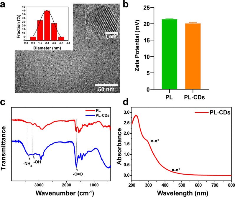
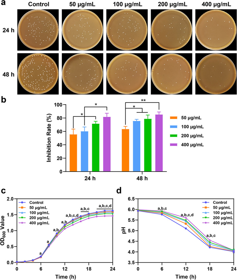
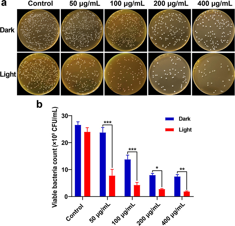
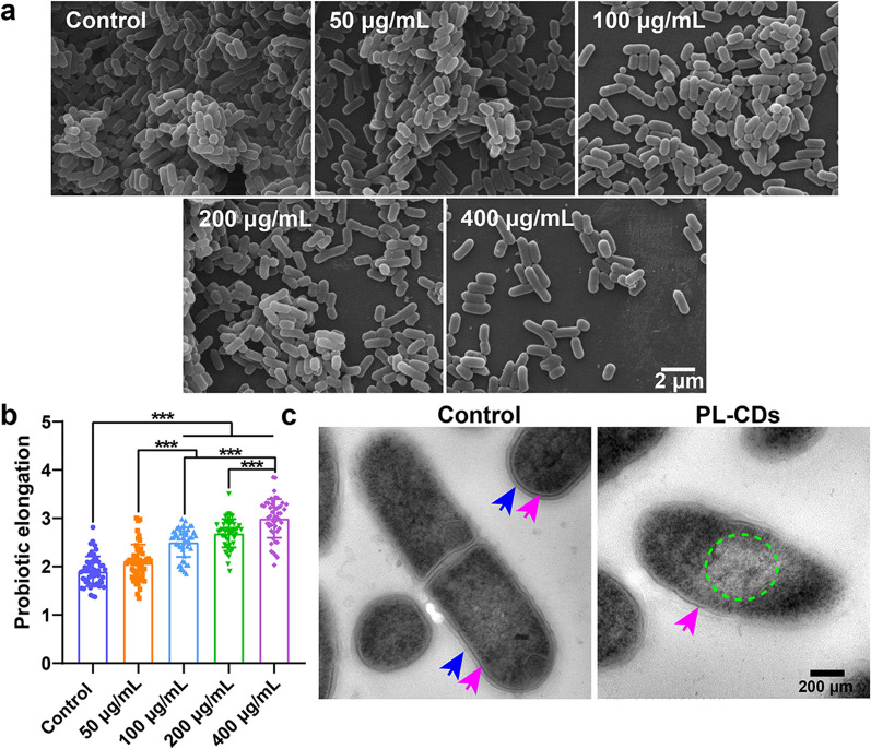
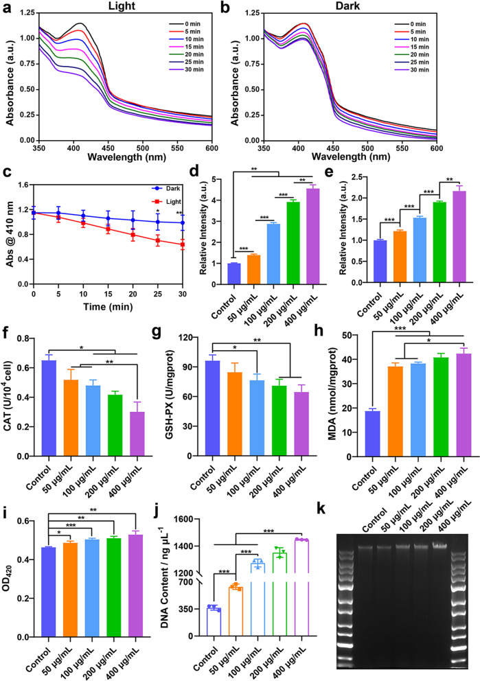
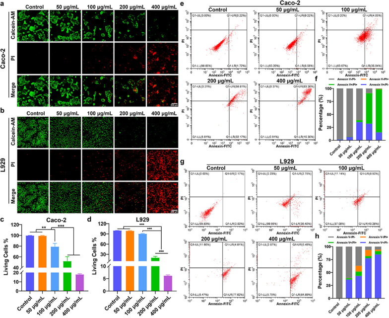
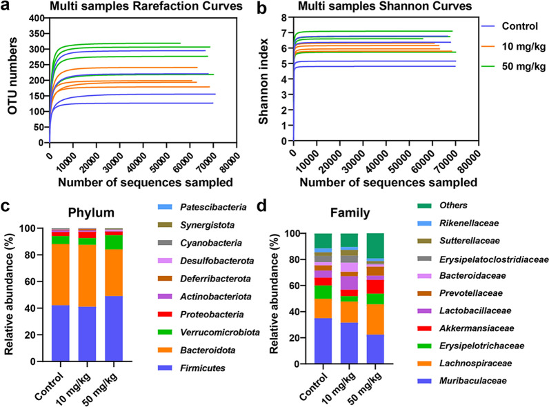
Results: Herein, CDs extracted from ε-poly-L-lysine (PL) were chosen to explore the modulation effect of CDs on probiotic behavior in vitro and intestinal remodeling in vivo. Results verify that PL-CDs negatively regulate Lactobacillus rhamnosus (L. rhamnosus) growth via increasing reactive oxygen species (ROS) production and reducing the antioxidant activity, which subsequently destroys membrane permeability and integrity. PL-CDs are also inclined to inhibit cell viability and accelerate cell apoptosis. In vivo, the gavage of PL-CDs is verified to induce inflammatory infiltration and barrier damage in mice. Moreover, PL-CDs are found to increase the Firmicutes to Bacteroidota (F/B) ratio and the relative abundance of Lachnospiraceae while decreasing that of Muribaculaceae.
Conclusion: Overall, these evidences indicate that PL-CDs may inevitably result in intestinal flora dysbiosis via inhibiting probiotic growth and simultaneously activating intestinal inflammation, thus causing pathological damage to the intestine, which provides an effective and insightful reference for the potential risk of CDs from the perspective of intestinal remodeling.
Keywords: Carbon dots; Inflammation; Intestinal flora; Probiotics; Reactive oxygen species.
© 2023. The Author(s).
Conflict of interest statement
The authors declare no competing interests.
Figures


Similar articles
-
Natural medicines-derived carbon dots as novel oral antioxidant administration strategy for ulcerative colitis therapy.J Nanobiotechnology. 2024 Aug 27;22(1):511. doi: 10.1186/s12951-024-02702-2. J Nanobiotechnology. 2024. PMID: 39187876 Free PMC article.
-
Lacticaseibacillus rhamnosus TR08 alleviated intestinal injury and modulated microbiota dysbiosis in septic mice.BMC Microbiol. 2021 Sep 18;21(1):249. doi: 10.1186/s12866-021-02317-9. BMC Microbiol. 2021. PMID: 34536996 Free PMC article.
-
Effects of Intestinal Microorganisms on Influenza-Infected Mice with Antibiotic-Induced Intestinal Dysbiosis, through the TLR7 Signaling Pathway.Front Biosci (Landmark Ed). 2023 Mar 2;28(3):43. doi: 10.31083/j.fbl2803043. Front Biosci (Landmark Ed). 2023. PMID: 37005752
-
Is intestinal inflammation linking dysbiosis to gut barrier dysfunction during liver disease?Expert Rev Gastroenterol Hepatol. 2015;9(8):1069-76. doi: 10.1586/17474124.2015.1057122. Epub 2015 Jun 18. Expert Rev Gastroenterol Hepatol. 2015. PMID: 26088524 Free PMC article. Review.
-
Probiotics and Probiotic-Derived Functional Factors-Mechanistic Insights Into Applications for Intestinal Homeostasis.Front Immunol. 2020 Jul 3;11:1428. doi: 10.3389/fimmu.2020.01428. eCollection 2020. Front Immunol. 2020. PMID: 32719681 Free PMC article. Review.
Cited by
-
The mechanism of tea tree oil regulating the damage of hydrogen sulfide to spleen and intestine of chicken.Poult Sci. 2025 Jan;104(1):104605. doi: 10.1016/j.psj.2024.104605. Epub 2024 Nov 28. Poult Sci. 2025. PMID: 39626606 Free PMC article.
-
Ozone controls the metabolism of tryptophan protecting against sepsis-induced intestinal damage by activating aryl hydrocarbon receptor.World J Gastroenterol. 2025 May 7;31(17):105411. doi: 10.3748/wjg.v31.i17.105411. World J Gastroenterol. 2025. PMID: 40521273 Free PMC article.
-
Recent progress in carbon dots for anti-pathogen applications in oral cavity.Front Cell Infect Microbiol. 2023 Sep 15;13:1251309. doi: 10.3389/fcimb.2023.1251309. eCollection 2023. Front Cell Infect Microbiol. 2023. PMID: 37780847 Free PMC article. Review.
-
A dynamic online nomogram for predicts delayed postoperative bleeding after colorectal polyp surgery.Sci Rep. 2024 Aug 25;14(1):19728. doi: 10.1038/s41598-024-70635-9. Sci Rep. 2024. PMID: 39183349 Free PMC article.
-
Ligilactobacillus salivarius LZZAY01 accelerated autophagy and apoptosis in colon cancer cells and improved gut microbiota in CAC mice.Microbiol Spectr. 2025 Feb 4;13(2):e0186124. doi: 10.1128/spectrum.01861-24. Epub 2025 Jan 10. Microbiol Spectr. 2025. PMID: 39792005 Free PMC article.
References
-
- Yao B, Huang H, Liu Y, Kang Z. Carbon dots: a small conundrum. Trends Chem. 2019;1:235–246. doi: 10.1016/j.trechm.2019.02.003. - DOI
-
- Li P, Han F, Cao W, Zhang G, Li J, Zhou J, et al. Carbon quantum dots derived from lysine and arginine simultaneously scavenge bacteria and promote tissue repair. Appl Mater Today. 2020;19:100601. doi: 10.1016/j.apmt.2020.100601. - DOI
MeSH terms
Substances
Grants and funding
- 31900957/National Natural Science Foundation of China
- 82173495/National Natural Science Foundation of China
- 2019KJE015/Innovation and technology program for the excellent youth scholars of higher education of Shandong province
- 2021Q069/Traditional Chinese Medicine Science and Technology Project of Shandong province
- KTRDHA-Y201902/Open Fund of Tianjin Enterprise Key Laboratory for Application Research of Hyaluronic Acid
LinkOut - more resources
Full Text Sources
