

Apical pulmonary lesions suspected of malignancy visible on neck CT angiography performed for acute stroke: Prevalence, treatment, and clinical implications - the PLEURA study
- PMID: 37231698
- PMCID: PMC10334179
- DOI: 10.1177/23969873231151488
Apical pulmonary lesions suspected of malignancy visible on neck CT angiography performed for acute stroke: Prevalence, treatment, and clinical implications - the PLEURA study
Abstract
Background: Computed tomography angiography (CTA) of the supraaortic arteries is commonly used for acute stroke workup and may reveal apical pulmonary lesions (APL).
Aim: To determine the prevalence, follow-up algorithms, and in-hospital outcomes of stroke patients with APL on CTA.
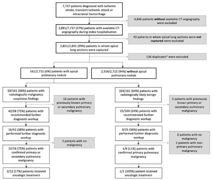
Methods: We retrospectively included consecutive adult patients with ischemic stroke, transient ischemic attack, or intracerebral hemorrhage and available CTA at a tertiary hospital between January 2014 and May 2021. We reviewed all CTA reports for the presence of APL. APL were classified as malignancy suspicious or benign appearing based on radiological-morphological criteria. We performed regression analyses to investigate the impact of malignancy suspicious APL on different in-hospital outcome parameters.
Results: Among 2715 patients, APL on CTA were found in 161 patients (5.9% [95%CI: 5.1-6.9]; 161/2715). Suspicion of malignancy was present in one third of patients with APL (36.0% [95%CI: 29.0-43.7]; 58/161), 42 of whom (72.4% [95%CI: 60.0-82.2]; 42/58) had no history of lung cancer or metastases. When performed, further investigations confirmed primary or secondary pulmonary malignancy in three-quarters (75.0% [95%CI: 50.5-89.8]; 12/16), with two patients (16.7% [95%CI: 4.7-44.8]; 2/12) receiving de novo oncologic therapy. In multivariable regression, the presence of radiologically malignancy suspicious APL was associated with higher NIHSS scores at 24 h (beta = 0.67, 95%CI: 0.28-1.06, p = 0.001) and all-cause in-hospital mortality (aOR = 3.83, 95%CI: 1.29-9.94, p = 0.01).
Conclusions: One in seventeen patients shows APL on CTA, of which one-third is malignancy suspicious. Further work-up confirmed pulmonary malignancy in a substantial number of patients triggering potentially life-saving oncologic therapy.
Keywords: CT angiography; Stroke; cancer; pulmonary lesions.
Conflict of interest statement
The author(s) declared the following potential conflicts of interest with respect to the research, authorship, and/or publication of this article: MP has received funds from Medtronic, Stryker, Penumbra, Phenox, Rapid Medical. He has received speakers honoraria from Stryker, Penumbra, Acandis, Phenox, Medtronic. GMDM received speaker honoraria from Medtronic; he declares no other conflicts of interest related to this work. The remaining authors report no conflicts of interest related to this work.
Figures
References
-
- MacMahon H, Naidich DP, Goo JM, et al.. Guidelines for management of incidental pulmonary nodules detected on CT images: from the fleischner society 2017. Radiology 2017; 284: 228–243. - PubMed
-
- Doyle SJ, George BP, Holloway RG, et al.. Incidental findings in radiographic imaging for inpatients with acute ischemic stroke. J Stroke Cerebrovasc Dis 2018; 27: 3131–3136. - PubMed
-
- Bentsen L, Christensen A, Havsteen I, et al.. Frequency of new pulmonary neoplasm incidentally detected by computed tomography angiography in acute stroke patients-a single-center study. J Stroke Cerebrovasc Dis 2015; 24: 1008–1012. - PubMed
-
- Verschoof MA, Groot AE, de Bruijn S, et al.. Clinical Outcome after endovascular treatment in patients with active cancer and ischemic stroke: a MR CLEAN registry substudy. Neurology 2022; 98: e993–e1001. - PubMed
Publication types
MeSH terms
LinkOut - more resources
Full Text Sources
Medical
