Review
. 2024;46(1):1-21.
doi: 10.1159/000530898. Epub 2023 May 10.

Out of Line or Altered States? Neural Progenitors as a Target in a Polygenic Neurodevelopmental Disorder

Affiliations
Review

Out of Line or Altered States? Neural Progenitors as a Target in a Polygenic Neurodevelopmental Disorder

Shah Rukh et al. Dev Neurosci. 2024.

Abstract

The genesis of a mature complement of neurons is thought to require, at least in part, precursor cell lineages in which neural progenitors have distinct identities recognized by exclusive expression of one or a few molecular markers. Nevertheless, limited progenitor types distinguished by specific markers and lineal progression through such subclasses cannot easily yield the magnitude of neuronal diversity in most regions of the nervous system. The late Verne Caviness, to whom this edition of Developmental Neuroscience is dedicated, recognized this mismatch. In his pioneering work on the histogenesis of the cerebral cortex, he acknowledged the additional flexibility required to generate multiple classes of cortical projection and interneurons. This flexibility may be accomplished by establishing cell states in which levels rather than binary expression or repression of individual genes vary across each progenitor's shared transcriptome. Such states may reflect local, stochastic signaling via soluble factors or coincidence of cell surface ligand/receptor pairs in subsets of neighboring progenitors. This probabilistic, rather than determined, signaling could modify transcription levels via multiple pathways within an apparently uniform population of progenitors. Progenitor states, therefore, rather than lineal relationships between types may underlie the generation of neuronal diversity in most regions of the nervous system. Moreover, mechanisms that influence variation required for flexible progenitor states may be targets for pathological changes in a broad range of neurodevelopmental disorders, especially those with polygenic origins.

Keywords: 22q11 deletion syndrome; Animal model; Cell lineage; Cerebral cortex; Neural development; Neurogenesis.


Conflict of interest statement

Conflict of Interest Statement

None of the authors have conflicts of interest to declare.

Figures

Fig. 1.
Fig. 1.
Neural progenitor specification and its relationship with CNS and PNS neuronal diversity. a Neuronal diversity may depend upon progenitor identities (distinct colors in dividing cells) that determine differentiation capacity of postmitotic progeny (related colors, top). Extrinsic signals may establish or reinforce discrete progenitor identities (multicolored arrows) during lineage progression. b Neuronal diversity may be independent of lineage, relying primarily upon combinations of extrinsic signals (multicolor arrows) that act on postmitotic neuroblasts to establish distinct identities. c Current views of lineage progression in the cerebral cortex. Radial glia (apical progenitors [AP]) in the ventricular zone ([VZ], adjacent to the cerebral ventricles [V]) primarily generate projection neurons (PNs) in layers 5/6 early in cortical histogenesis. They also act as guides for postmitotic neuroblasts (NBs) as they migrate radially to their laminar positions. At later stages, radial glia generate basal progenitors (BPs) which diversify in the subventricular zone (SVZ) to include basal radial glia (blue cell with process). BPs generate primarily layer 2/3 PNs. d Trigeminal ganglion progenitor diversity and presumed lineages. Two distinct neural crest progenitor types: one from Wnt1-expressing neuroepithelial neural tube progenitors (green) and another from non-Wnt1 neural tube progenitors (blue), migrate and coalesce with trigeminal cranial placode-derived progenitors (red). These populations, with distinct derivations, give rise, respectively, to mechanosensory (red) and nociceptive neurons (green and blue) axons extending into the distal and proximal branches of the trigeminal nerve (c after [60, 61]; d based upon data from [16, 62]).
Fig. 2.
Fig. 2.
Multiple contiguous mouse orthologues on mouse chromosome 16 of genes that define the region of human chromosome 22 deleted in 22q11.2 deletion syndrome are selectively expressed in the VZ/SVZ and trigeminal ganglion during neurogenesis. Top: a schematic of the region of mouse chromosome 16 where 28 colinear orthologues of the genes from the minimal critical deleted region associated with 22q11DS are located. Gray bars indicate the loci of protein-coding genes. Several 22q11 genes are expressed selectively in the developing cortical mantle (top, each panel) as well as the trigeminal ganglion (CNgV; bottom, each panel) during cortical histogenesis or gangliogenesis. Lines extend from protein-coding loci on the chromosomal map to identify images of cortical and CNgV expression patterns of the encoded transcript. Expression of a subset of 22q11 genes (Dgcr8, Ufd1, Ranbp1, Comt, Cldn5, Trmt2a, Txnrd2, Cldn5, Cdc45) is selectively enhanced in the VZ/SVZ (inset, top right, each panel). The color code (bottom) indicates the diverse functional classes to which each of these 22q11 genes localized to the VZ/SVZ and CNgV belong (top, after [75]; images from Genepaint; functional category key from [124]).
Fig. 3.
Fig. 3.
Heterozygous deletion at human chromosome 22q11 and parallel heterozygous deletion of contiguous murine orthologues on mouse chromosome 16 results in behavioral disruption and cellular pathology associated with neurodevelopmental disorders (NDDs). Top left: the location of the heterozygous deletion proximal to the centromere on the q arm of chromosome 22 that is associated with 22q11DS. Top right: the phenotypic spectrum of 22q11DS includes substantially elevated frequency of behavioral disorders that parallel clinically defined NDDs including ADHD, autism spectrum disorders (ASD), schizophrenia (Scz), and anxiety disorder. Functional imaging studies of individuals with 22q11DS indicate cortical circuit dysfunction, and structural imaging as well as a limited postmortem analyses have found likely foci of cellular brain pathology. In addition to clinically diagnosed NDDs associated with cortical circuitry and function, infants and toddlers with 22q11DS have perinatal dysphagia – difficulties with suckling, feeding, and swallowing – followed by oromotor difficulties including aberrant food ingestion, swallowing, and speech throughout the lifespan. Bottom left: location of heterozygous deletion of contiguous 22q11 orthologues that define the minimal critical deleted region associated with 22q11DS on mouse chromosome 16. Bottom right: although it is not possible to identify behavioral states in mice that fully parallel those in ADHD, ASD, Scz, or anxiety disorder, LgDel mice with a deletion on mmChr16 orthologous to that on hChr 22 have a spectrum of behavioral deficits and changes in cortical as well as peripheral neural circuits that can be related to aspects of human clinical disorders seen in individuals with 22q11DS throughout the lifespan (figure modified from [75]).
Fig. 4.
Fig. 4.
CNS and PNS neural progenitors are compromised by heterozygous deletion of murine orthologues of human 22q11 genes. a Selective disruption of basal progenitor proliferation during cortical histogenesis by heterozygous deletion of murine 22q11 orthologues. Top right: in E13.5 LgDel fetuses, as the initial population of basal progenitors is being generated, there is a diminished frequency of Tbr2+ basal progenitors that can also be labeled acutely with BrdU, indicating active proliferation. Bottom right: in neonatal LgDel mice (postnatal day P8), the frequency of layer 2/3 PNs, labeled selectively for Cux1 as well as the general neuronal marker NeuN (RbFox3), declines significantly; however, the frequency of NeuN+ or Ctip2+ neurons (a selective marker for layer 5/6 PNs) in layer 5/6 is not substantially changed. b Disrupted neurogenesis and progenitor proliferation during trigeminal ganglion (CNgV) differentiation due to heterozygous deletion of murine 22q11 orthologues. Top panels: there is an apparent increase in the frequency of newly generated neurons, labeled with NeuN, in the E10.5 LgDel CNgV. Middle panels: increased frequency of early generated CNgV neurons in LgDel apparently reflects increased symmetric neuron-neuron as well as increased asymmetric precursor-neuron divisions of neural crest-associated (Sox2+) CNgV neural progenitors. These data were generated using a pair-cell assay in which CNgV cells are dissociated, plated in microwells at very low density, and allowed to divide for 21 h. Isolated pairs of cells are presumed to derive from individual progenitors, and their identities are then probed with specific progenitor markers (in this case Sox2) as well as neuronal markers (in this case, βIII-tubulin). Bottom panels: the apparent frequency of subclasses of TrpV1+ nociceptive sensory neurons, particularly those labeled for TrpV1 and via recombination driven by Wnt1 to mark a subpopulation of neural crest-derived progenitors at earlier stages, increases in P8 LgDel CNgV. This increase is paralleled by increased abundance of TrpV1 mRNA in LgDel versus WT, measured by qPCR in samples of dissected P8 CNgV (a adapted from [3]; b from [16]).
Fig. 5.
Fig. 5.
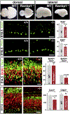
Homozygous null mutation of the 22q11 deleted gene Ranbp1 results in microcephaly due to diminished apical and basal progenitor proliferation. Top panels: at E18.5, Ranbp1−/− mice, who die at birth, are visibly microcephalic, based upon head size (not shown) and brain size, especially that of the cerebral hemispheres. These changes in brain size are visible both from a dorsal (left) and latral (right) view. Middle panels: at E10.5, when the cortical rudiment is still a proliferative neurepithelium with a small number of postmitotic neurons, active proliferation of neuroepithelial/apical progenitors, labeled with Ki67, is diminished in Ranbp1−/− fetuses, as is the frequency of PH3+ presumed mitotic progenitors. Lower panels: by E14, when basal progenitor genesis from apical progenitors has begun, there are fewer Tbr2+ basal progenitors acutely labeled by BrdU in Ranbp1/ fetuses. By E17.5, when an appreciable number of layer 2/3 PNs, labeled here by Cux1, have accumulated in the WT cortex, there are significantly fewer Cux1+ layer 2/3 PNs in the Ranbp1−/− cortex; however, the frequency of Ctip2+ layer 5/6 PNs is not significantly reduced (all panels adapted from [15]).
Fig. 6.
Fig. 6.
Transcriptional divergence of E10.5 LgDel CNgV neural progenitors and newly generated neurons. Top panels: we dissected CNgV from WT and LgDel E10.5 fetuses, an age when substantial numbers of placodal (Six1+, red) and neural crest (Wnt1Cre-recombined + or −, green and blue, respectively) can be seen in the ganglion (far right). These dissected CNgV from multiple embryos and multiple litters were pooled to collect 5 biological replicates for RNAseq transcriptome analysis. Middle panels: comparison of the 5 biological replicate CNgV transcriptomes identified 134 genes that are differentially expressed in LgDel versus WT, based upon a significance threshold of FDR q < 0.1 (left). Of these 134 genes, 38 can be informatically identified as potential targets for transcriptional regulation by Six1, a diagnostic transcription factor expressed in placode-derived CNgV progenitors, and 130 can be identified as potential targets for transcriptional regulation by Sox10, a diagnostic transcription factor marker for a subpopulation of neural crest-derived CNgV progenitors (right). Lower panels: there is a significant transcriptome-wide increase in the coefficient of variation for genes expressed in the E10.5 LgDel versus WT CNgV. Increased variation is seen for 22q11 genes whose expression is detected in the E10.5 CNgV (red dots) as well as the 134 genes differentially expressed between LgDel and WT (orange dots). Increased variation can be seen across functional categories (right) that include fundamental cellular metabolic and protein/membrane trafficking processes (amino acid synthesis and Golgi) as well as those specific for neural progenitors and differentiating neurons (proneural genes, axon extension genes). Asterisks indicate significance levels as shown in the top left corner (all panels adapted from [123]).
Fig. 7.
Fig. 7.
State-dependent regulation and diversification of neural progenitor classes whose broad identities are defined by molecular markers. These progenitors, of which two examples are shown here in detail, retain the unique expression of their diagnostic molecular marker, often reflecting derivation from another broad class of progenitors. Their transcriptional identities (indicated here by the colored bars on the histograms within the nuclei of the two cells) are at one level of analysis – absolute presence of multiple transcripts – equivalent. Their states, however, are divergent based upon variation of levels of expression of the set of transcripts shared by the two otherwise indistinguishable progenitors. These states, indicated here based upon levels of transcription of identified genes, are likely established by diffusible extrinsic signals as well as signaling that relies upon cell-cell contact. The targets of these signals for modifying progenitor state likely include activation or repression of signaling pathways that influence transcription as well as chromatin regulation via direct DNA methylation or changes in histone modifications. NDD pathogenic processes may target these state-modulation mechanisms without changing broad identities of molecular marker-defined progenitor classes. The consequences of these “altered states” would be recognized as increases or decreases in the transcription variability due to quantitative alteration of signaling pathways and their downstream targets including posttranslational modification of transcriptional regulators as well as chromatin-modifying enzymes.

Similar articles

References

    1. Rakic P, Caviness VS Jr. Cortical development: view from neurological mutants two decades later. Neuron. 1995. Jun;14(6):1101–4. - PubMed
    1. Caviness VS Jr, Rakic P. Mechanisms of cortical development: a view from mutations in mice. Annu Rev Neurosci. 1978;1:297–326. - PubMed
    1. Meechan DW, Tucker ES, Maynard TM, LaMantia AS. Diminished dosage of 22q11 genes disrupts neurogenesis and cortical development in a mouse model of 22q11 deletion/DiGeorge syndrome. Proc Natl Acad Sci U S A. 2009. Sep 22;106(38):16434–45. - PMC - PubMed
    1. Maynard TM, Haskell GT, Peters AZ, Sikich L, Lieberman JA, LaMantia AS. A comprehensive analysis of 22q11 gene expression in the developing and adult brain. Proc Natl Acad Sci U S A. 2003. Nov 25;100(24):14433–8. - PMC - PubMed
    1. Herrup K. Role of staggerer gene in determining cell number in cerebellar cortex. I. Granule cell death is an indirect consequence of staggerer gene action. Brain Res. 1983. Dec;313(2):267–74. - PubMed

Publication types

LinkOut - more resources