

Oogenesis in Caenorhabditis elegans
- PMID: 37232019
- PMCID: PMC10659005
- DOI: 10.1159/000531019
Oogenesis in Caenorhabditis elegans
Abstract
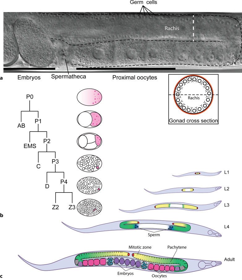
Background: The nematode, Caenorhabditis elegans has proven itself as a valuable model for investigating metazoan biology. C. elegans have a transparent body, an invariant cell lineage, and a high level of genetic conservation which makes it a desirable model organism. Although used to elucidate many aspects of somatic biology, a distinct advantage of C. elegans is its well annotated germline which allows all aspects of oogenesis to be observed in real time within a single animal. C. elegans hermaphrodites have two U-shaped gonad arms which produce their own sperm that is later stored to fertilise their own oocytes. These two germlines take up much of the internal space of each animal and germ cells are therefore the most abundant cell present within each animal. This feature and the genetic phenotypes observed for mutant worm gonads have allowed many novel findings that established our early understanding of germ cell dynamics. The mutant phenotypes also allowed key features of meiosis and germ cell maturation to be unveiled.
Summary: This review will focus on the key aspects that make C. elegans an outstanding model for exploring each feature of oogenesis. This will include the fundamental steps associated with germline function and germ cell maturation and will be of use for those interested in exploring reproductive metazoan biology.
Key messages: Since germ cell biology is highly conserved in animals, much can be gained from study of a simple metazoan like C. elegans. Past findings have enhanced understanding on topics that would be more laborious or challenging in more complex animal models.
Keywords: Caenorhabditis elegans; Development; Oocyte meiotic maturation; Oogenesis.
© 2023 The Author(s). Published by S. Karger AG, Basel.
Conflict of interest statement
The authors have no conflicts of interest.
Figures
Similar articles
-
SACY-1 DEAD-Box helicase links the somatic control of oocyte meiotic maturation to the sperm-to-oocyte switch and gamete maintenance in Caenorhabditis elegans.Genetics. 2012 Nov;192(3):905-28. doi: 10.1534/genetics.112.143271. Epub 2012 Aug 10. Genetics. 2012. PMID: 22887816 Free PMC article.
-
Germ cell survival in C. elegans and C. remanei is affected when the DEAD box RNA helicases VBH-1 or Cre-VBH-1 are silenced.Genesis. 2012 Nov;50(11):801-18. doi: 10.1002/dvg.22043. Epub 2012 Jul 11. Genesis. 2012. PMID: 22674898
-
The Caenorhabditis elegans homologue of deleted in azoospermia is involved in the sperm/oocyte switch.Mol Biol Cell. 2006 Jul;17(7):3147-55. doi: 10.1091/mbc.e05-11-1067. Epub 2006 Apr 26. Mol Biol Cell. 2006. PMID: 16641369 Free PMC article.
-
Start me up: cell signaling and the journey from oocyte to embryo in C. elegans.Dev Dyn. 2006 Mar;235(3):571-85. doi: 10.1002/dvdy.20662. Dev Dyn. 2006. PMID: 16372336 Review.
-
Control of germ cell differentiation in Caenorhabditis elegans.Ciba Found Symp. 1994;182:179-88; discussion 189-92. doi: 10.1002/9780470514573.ch10. Ciba Found Symp. 1994. PMID: 7835149 Review.
Cited by
-
EYA-1 is required for genomic integrity independent of H2AX signalling in Caenorhabditis elegans.Mol Biol Rep. 2024 Sep 24;51(1):1009. doi: 10.1007/s11033-024-09933-4. Mol Biol Rep. 2024. PMID: 39316168 Free PMC article.
-
Host environment shapes filarial parasite fitness and Wolbachia endosymbionts dynamics.PLoS Pathog. 2025 Jul 11;21(7):e1013301. doi: 10.1371/journal.ppat.1013301. eCollection 2025 Jul. PLoS Pathog. 2025. PMID: 40644522 Free PMC article.
References
Publication types
MeSH terms
Substances
LinkOut - more resources
Full Text Sources
Miscellaneous
