Review
. 2023 Jul;54(7):1930-1942.
doi: 10.1161/STROKEAHA.123.040205. Epub 2023 May 26.

Haptoglobin Treatment for Aneurysmal Subarachnoid Hemorrhage: Review and Expert Consensus on Clinical Translation

Collaborators, Affiliations
Review

Haptoglobin Treatment for Aneurysmal Subarachnoid Hemorrhage: Review and Expert Consensus on Clinical Translation

Ian Galea et al. Stroke. 2023 Jul.

Abstract

Aneurysmal subarachnoid hemorrhage (aSAH) is a devastating form of stroke frequently affecting young to middle-aged adults, with an unmet need to improve outcome. This special report focusses on the development of intrathecal haptoglobin supplementation as a treatment by reviewing current knowledge and progress, arriving at a Delphi-based global consensus regarding the pathophysiological role of extracellular hemoglobin and research priorities for clinical translation of hemoglobin-scavenging therapeutics. After aneurysmal subarachnoid hemorrhage, erythrocyte lysis generates cell-free hemoglobin in the cerebrospinal fluid, which is a strong determinant of secondary brain injury and long-term clinical outcome. Haptoglobin is the body's first-line defense against cell-free hemoglobin by binding it irreversibly, preventing translocation of hemoglobin into the brain parenchyma and nitric oxide-sensitive functional compartments of cerebral arteries. In mouse and sheep models, intraventricular administration of haptoglobin reversed hemoglobin-induced clinical, histological, and biochemical features of human aneurysmal subarachnoid hemorrhage. Clinical translation of this strategy imposes unique challenges set by the novel mode of action and the anticipated need for intrathecal drug administration, necessitating early input from stakeholders. Practising clinicians (n=72) and scientific experts (n=28) from 5 continents participated in the Delphi study. Inflammation, microvascular spasm, initial intracranial pressure increase, and disruption of nitric oxide signaling were deemed the most important pathophysiological pathways determining outcome. Cell-free hemoglobin was thought to play an important role mostly in pathways related to iron toxicity, oxidative stress, nitric oxide, and inflammation. While useful, there was consensus that further preclinical work was not a priority, with most believing the field was ready for an early phase trial. The highest research priorities were related to confirming haptoglobin's anticipated safety, individualized versus standard dosing, timing of treatment, pharmacokinetics, pharmacodynamics, and outcome measure selection. These results highlight the need for early phase trials of intracranial haptoglobin for aneurysmal subarachnoid hemorrhage, and the value of early input from clinical disciplines on a global scale during the early stages of clinical translation.

Keywords: blood; haptoglobins; hemoglobins; subarachnoid hemorrhage; therapeutics.


Conflict of interest statement

Disclosures Dr Galea and D. Bulters declare research funding and consulting fees from BioProducts Laboratory Limited and Evgen Pharma. Dr Galea received research funding related to haptoglobin from the Medical Research Council and Engineering and Physical Sciences Research Council. Drs Galea and Hugelshofer disclose consulting fees from CSL Behring. Drs Schaer and Hugelshofer received research funding from CSL Behring and are inventors on patents related to the use of haptoglobin.

Figures

Figure 1.
Figure 1.
Root triggers. Acute intracranial pressure rise and cell-free hemoglobin as root triggers leading to downstream causes of brain injury post-aneurysmal subarachnoid hemorrhage.
Figure 2.
Figure 2.
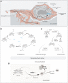
Mechanisms of Hb toxicity. A, Hemoglobin dimers in the subarachnoid space can penetrate: (1) the interstitial space between smooth muscle cells of cerebral arteries. Consumption of endothelium-derived nitric oxide causes vasospasm; (2) cortical tissue, promoting oxidative damage and inflammation. B, Reactions of NO with Hb across a range of O2 liganded [oxy-Hb(Fe2+O2)] and nonliganded states [deoxy-Hb, Hb(Fe2+) and met-Hb [Hb(Fe3+)]. The first NO reaction with oxy-Hb produces Hb(Fe3+) and nitrate (NO3). A second, slower NO consumption step reaction is proposed to involve a series of reaction intermediates, which ultimately react with water and production of nitrite (NO2), H+, and Hb(Fe2+). In a third reaction, NO binds to Hb(Fe2+). C, Oxidized Hb(Fe3+) releases heme, which promotes radical reactions and inflammation via TLR receptor signaling. D, In macrophages, heme is metabolized by heme oxygenase (HMOX-1) generating carbon monoxide (CO), bilirubin, and iron. Iron is either exported or stored in ferritin.
Figure 3.
Figure 3.
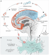
Haptoglobin treatment. A, SAH after aneurysmal rupture forms a blood clot. Erythrolysis releases hemoglobin tetramers, which dissociate into dimers in CSF (red in the online version). Small Hb dimers penetrate the brain parenchyma and NO-sensitive arterial compartments to cause secondary brain injury. Therapeutic haptoglobin (blue in the online version) administered via an intrathecal catheter distributes throughout the cerebrospinal fluid (CSF) compartment and binds free hemoglobin. The large hemoglobin-haptoglobin complex remains confined outside the parenchyma and vulnerable arterial compartments, thereby protecting from hemoglobin-induced damage. The hemoglobin-haptoglobin complex is cleared by physiological drainage pathways and drained through intraventricular and/or lumbar catheters (Figure S1). B, The role of macrophages in erythrophagocytosis and hemoglobin-haptoglobin complex clearance. Following degradation, heme is metabolized to bilirubin, carbon monoxide, and iron through heme-mediated induction of HMOX-1 (heme-oxygenase 1). Heme-induced activation of NRF2 (nuclear factor erythroid 2-related factor 2) signaling induces an anti-inflammatory macrophage phenotype (ie, erythrophagocyte).
Figure 4.
Figure 4.
Delphi study participants. Geographic distribution and demographics. Please note that clinical disciplines add up to more than 100% since some had dual expertise.
Figure 5.
Figure 5.
Delphi study results. A, Change in responses between Delphi rounds (***P<0.001, Wilcoxon). B, Clinicians and scientists were asked to rank 10 pathophysiological pathways according to their role in secondary brain injury (SBI) and estimate the role of hemoglobin for each pathway using a Likert scale with a scoring range from 1 (extremely important) to 5 (not sure). The plots show the kernel density estimation of participants’ responses. The solid line connects the group medians. C and D, Scientists (C) and clinicians (D) ranked potential research questions according to their priority and estimated the importance of each research question with a Likert system with a scoring range from 1 (extremely important) to 9 (extremely unimportant). The plots show the kernel density estimation of participants’ responses. The solid line connects the group medians. CSF indicates cerebrospinal fluid.

References

    1. Etminan N, Chang H-S, Hackenberg K, de Rooij NK, Vergouwen MDI, Rinkel GJE, Algra A. Worldwide incidence of aneurysmal subarachnoid hemorrhage according to region, time period, blood pressure, and smoking prevalence in the population. Jama Neurol. 2019;76:588–597. doi: 10.1001/jamaneurol.2019.0006 - PMC - PubMed
    1. Taylor TN, Davis PH, Torner JC, Holmes J, Meyer JW, Jacobson MF. Lifetime cost of stroke in the United States. Stroke. 1996;27:1459–1466. doi: 10.1161/01.str.27.9.1459 - PubMed
    1. Chou SH-Y. Subarachnoid hemorrhage. Continuum. 2021;27:1201–1245. doi: 10.1212/CON.0000000000001052 - PubMed
    1. Pickard JD, Murray GD, Illingworth R, Shaw MD, Teasdale GM, Foy PM, Humphrey PR, Lang DA, Nelson R, Richards P. Effect of oral nimodipine on cerebral infarction and outcome after subarachnoid hemorrhage. BMJ. 1989;298:636–642. doi: 10.1136/bmj.298.6674.636 - PMC - PubMed
    1. Khan AU, Dulhanty L, Vail A, Tyrrell P, Galea J, Patel HC. Impact of specialist neurovascular care in subarachnoid haemorrhage. Clin Neurol Neurosurg. 2015;133:55–60. doi: 10.1016/j.clineuro.2015.03.006 - PubMed

Publication types