

Tomosynthesis-Detected Architectural Distortions: Correlations between Imaging Characteristics and Histopathologic Outcomes
- PMID: 37233322
- PMCID: PMC10219079
- DOI: 10.3390/jimaging9050103
Tomosynthesis-Detected Architectural Distortions: Correlations between Imaging Characteristics and Histopathologic Outcomes
Abstract
Objective: to determine the positive predictive value (PPV) of tomosynthesis (DBT)-detected architectural distortions (ADs) and evaluate correlations between AD's imaging characteristics and histopathologic outcomes.
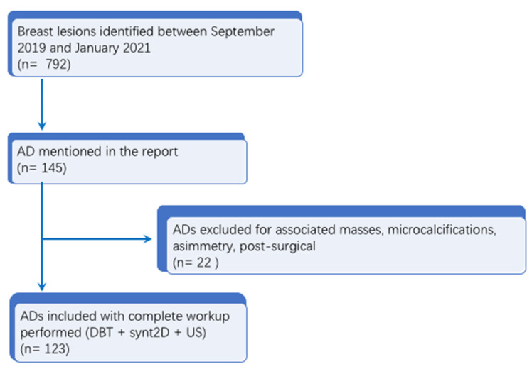
Methods: biopsies performed between 2019 and 2021 on ADs were included. Images were interpreted by dedicated breast imaging radiologists. Pathologic results after DBT-vacuum assisted biopsy (DBT-VAB) and core needle biopsy were compared with AD detected by DBT, synthetic2D (synt2D) and ultrasound (US).
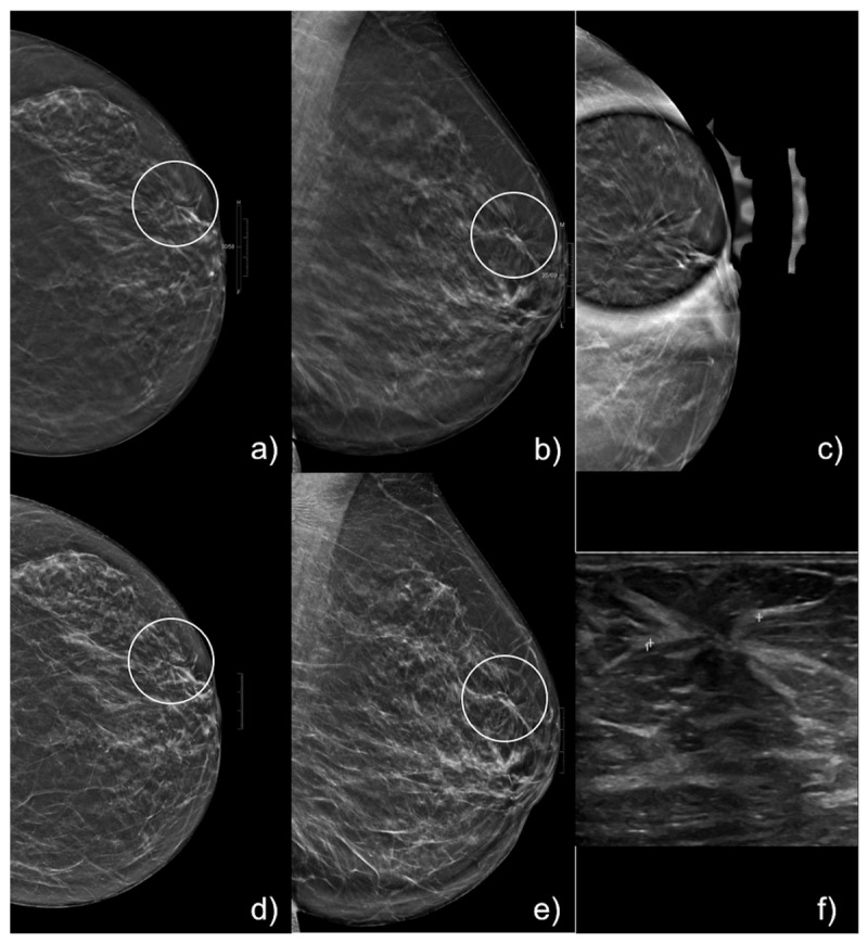
Results: US was performed to assess a correlation for ADs in all 123 cases and a US correlation was identified in 12/123 (9.7%) cases, which underwent US-guided core needle biopsy (CNB). The remaining 111/123 (90.2%) ADs were biopsied under DBT guidance. Among the 123 ADs included, 33/123 (26.8%) yielded malignant results. The overall PPV for malignancy was 30.1% (37/123). The imaging-specific PPV for malignancy was 19.2% (5/26) for DBT-only ADs, 28.2% (24/85) for ADs visible on DBT and synth2D mammography and 66.7% (8/12) for ADs with a US correlation with a statistically significant difference among the three groups (p = 0.01).
Conclusions: DBT-only ADs demonstrated a lower PPV of malignancy when compared with syntD mammography, and DBT detected ADs but not low enough to avoid biopsy. As the presence of a US correlate was found to be related with malignancy, it should increase the radiologist's level of suspicion, even when CNB returned a B3 result.
Keywords: B3 lesions; Breast Cancer Screening Program; DBT-vacuum assisted biopsy; architectural distortion; breast cancer; breast imaging; synthetic2D; tomosynthesis.
Conflict of interest statement
The authors declare no conflict of interest.
Figures
References
-
- D’Orsi C.J., Sickles E.A., Mendelson E.B., Morris E.A. ACR BI-RADS® Atlas, Breast Imaging Reporting and Data System. American College of Radiology; Reston, VA, USA: 2013.
-
- Babkina T.M., Gurando A.V., Kozarenko T.M., Gurando V.R., Telniy V.V., Pominchuk D.V. Detection of breast cancers represented as architectural distortion: A comparison of full-field digital mammography and digital breast tomosynthesis. Wiadomości Lek. 2021;74:1674–1679. doi: 10.36740/WLek202107121. - DOI - PubMed
LinkOut - more resources
Full Text Sources
Research Materials
