Review
. 2023 Apr 26;21(5):266.
doi: 10.3390/md21050266.

Diatom-Based Nanomedicine for Colorectal Cancer Treatment: New Approaches for Old Challenges

Affiliations
Review

Diatom-Based Nanomedicine for Colorectal Cancer Treatment: New Approaches for Old Challenges

Chiara Tramontano et al. Mar Drugs. .

Abstract

Colorectal cancer is among the most prevalent and lethal cancers globally. To address this emergency, countries have developed diffuse screening programs and innovative surgical techniques with a consequent decrease in mortality rates in non-metastatic patients. However, five years after diagnosis, metastatic CRC is still characterized by less than 20% survival. Most patients with metastatic CRC cannot be surgically treated. For them, the only option is treatment with conventional chemotherapies, which cause harmful side effects in normal tissues. In this context, nanomedicine can help traditional medicine overcome its limits. Diatomite nanoparticles (DNPs) are innovative nano-based drug delivery systems derived from the powder of diatom shells. Diatomite is a porous biosilica largely found in many areas of the world and approved by the Food and Drug Administration (FDA) for pharmaceutical and animal feed formulations. Diatomite nanoparticles with a size between 300 and 400 nm were shown to be biocompatible nanocarriers capable of delivering chemotherapeutic agents against specific targets while reducing off-target effects. This review discusses the treatment of colorectal cancer with conventional methods, highlighting the drawbacks of standard medicine and exploring innovative options based on the use of diatomite-based drug delivery systems. Three targeted treatments are considered: anti-angiogenetic drugs, antimetastatic drugs, and immune checkpoint inhibitors.

Keywords: biosilica; chemotherapy; colorectal cancer (CRC); diatoms; nanomedicine; nanotechnology; neoplasm metastasis.


Conflict of interest statement

The authors declare no conflict of interest.

Figures

Figure 1
Figure 1
Comparison between synthetic mesoporous silica (green, left) and diatomite silica (brown, right). Synthetic and natural silica show common advantages, such as a high surface area, tunable surface chemistry, and biocompatibility for drug delivery. Diatom biosilica must be converted into biodegradable silicon for therapeutic applications, while synthetic mesoporous silica is naturally excreted from the body. However, the production of NPs from diatom biosilica is cheaper, eco-friendly, and, overall, more convenient than its synthetic counterpart.
Figure 2
Figure 2
(a) SEM investigation of DEM-B12 interaction with HT29 cells mediated by the transcobalamin receptor. (b,c) Specific uptake of genetically engineered diatoms modified with the anti-CD20 antibody in cancer cells (b) and control cells (c). Scale bars are 50 µm (b,c). (df) Uptake of PSiNPs-DOX obtained from diatom biosilica in cancer cells. Cell nuclei were stained with DAPI (d), while the PSiNPs-DOX exhibited a natural red fluorescence (e). Merged channels are shown in (f). Scale bars are 25 µm (df). Adapted with permission from [43] (a), [47] (b,c), and [24] (df).
Figure 3
Figure 3
(a) Confocal microscopy on cells treated with siRNA*-modified diatomite nanovectors labeled with Dy547 (NPs). Cell nuclei and membranes were stained with Hoechst 33342 and WGA-Alexa Fluor 488, respectively. The scale bar is 20 μm. (b) A549 cells incubated for 24 h with PBS (control), bare frustules (AF), and Chi@AF-DOX at a concentration of 1 μg/mL (c) and 37 °C for 24 h. AOEB was used for staining the apoptotic bodies of cells, while green fluorescence represents viable cells and yellowish-orange cells represent apoptotic cells. (c) In vivo imaging results of IONP-embedded diatom accumulation in mice with (left) and without (right) the application of an external magnetic field. During the process, a higher signal (6.4 times stronger) of NP accumulation was observed in the tumors attached to a magnet. Reproduced and adapted with permission from [19] (a), [56] (b), and [38] (c).
Figure 4
Figure 4
Epithelial-to-mesenchymal transition (EMT) is driven by CRC cells’ transforming growth factor (TGF-β). Upregulation of TGF-β signaling promotes overexpression of mesenchymal markers, which favor EMT, migration, and invasion of CRC cells into a secondary tumor site. Anti-metastatic approaches include the blockade of the TGF-β-mediated pathway, inhibition of EMT, and promotion of a molecular and morphological cell transformation known as the mesenchymal-to-epithelial (MET) transition.
Figure 5
Figure 5
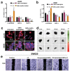
(a,b) Quantitative polymerase chain reaction (qPCR) analysis of EMT genes in LS-174T and SW620 cells incubated with 2.5 × 10⁻⁶ M LY, 50 μg mL−1 DNPs-AuNPs@Gel (empty), and DNPs-AuNPs-LY@Gel (releasing 2.5 × 10⁻⁶ M LY). Data were normalized to GAPDH expression and presented as fold change (FC) in gene expression relative to control. * p < 0.05, ** p < 0.005, *** p < 0.0005. (c) Representative images of DNPs-AuNPs-LY@Gel-mediated MET in LS-174T and SW620 cells. The scale bars are 100 μm. (d) Transmission electron microscopy (TEM) images of the encapsulated DNP dissolution at different pHs mimicking the GI. The scale bars are 300 nm. (e) Migration assay of SW620 cells incubated with 0.5% FBS DMEM (control), 2.5 µM LY, 26 µg mL−1 encapsulated DNPs (empty), and drug-loaded encapsulated DNPs in 0.5% FBS DMEM for 120 h. The scale bars are 500 µm. Adapted with permission from [57] (ac) and [35] (d).
Figure 6
Figure 6
(a) Confocal microscope images of A549 cells incubated with DNP–PNA* or PNA* for 24 h. The cell membrane was stained with CellMask Deep Red, while the cell nuclei were stained with DAPI. The images were acquired using a 63× objective. Scale bar is 50 µm. (b) DNP uptake kinetics in cancer cells though confocal (left) and Raman imaging (right) at different incubation times (6, 24, 72 h). (cf) Fluorescence-assisted cell sorting (FACWS) analysis of non-permeabilized A20 cells treated with PBS (control, c,e) or DNPs-siRNA (d,f). Adapted with permission from [59] (a), [104] (b), and [60] (cf).

References

    1. Ciardiello F., Ciardiello D., Martini G., Napolitano S., Tabernero J., Cervantes A. Clinical management of metastatic colorectal cancer in the era of precision medicine. CA Cancer J. Clin. 2022;72:372–401. doi: 10.3322/caac.21728. - DOI - PubMed
    1. Redmond E., Lally C., Cahill P. Chapter 3—Common Pathways in Cancer, Tumor Angiogenesis and Vascular Disease. In: Gottlieb R.A., Mehta P.K., editors. Cardio-Oncology. Academic Press; Boston, MA, USA: 2017. pp. 35–53. - DOI
    1. Compton C.C. Colorectal Carcinoma: Diagnostic, Prognostic, and Molecular Features. Mod. Pathol. 2003;16:376–388. doi: 10.1097/01.MP.0000062859.46942.93. - DOI - PubMed
    1. Gaffey M.J., Mills S.E., Lack E.E. Neuroendocrine Carcinoma of the Colon and Rectum A Clinicopathologic, Ultrastructural, and Immunohistochemical Study of 24 Cases. [(accessed on 21 April 2023)];Am. J. Surg. Pathol. 1990 14:1010–1023. doi: 10.1097/00000478-199011000-00003. Available online: https://journals.lww.com/ajsp/Fulltext/1990/11000/Neuroendocrine_Carcino.... - DOI - PubMed
    1. Nassar H., Ataya K., Hafez B., El Bsat A., Geagea L., Faraj W. Primary squamous cell carcinoma of the colon: A rare case report. Int. J. Surg. Case Rep. 2022;96:107383. doi: 10.1016/j.ijscr.2022.107383. - DOI - PMC - PubMed