Review
. 2023 May 21;13(5):675.
doi: 10.3390/metabo13050675.

Molecular Mechanisms of Western Diet-Induced Obesity and Obesity-Related Carcinogenesis-A Narrative Review

Affiliations
Review

Molecular Mechanisms of Western Diet-Induced Obesity and Obesity-Related Carcinogenesis-A Narrative Review

Dhruvi Lathigara et al. Metabolites. .

Abstract

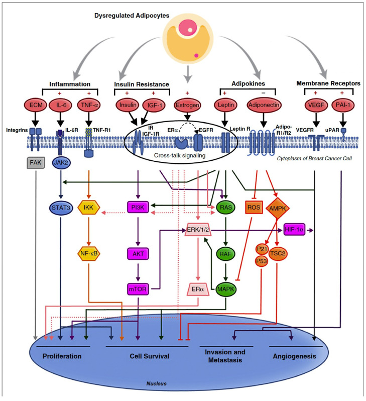
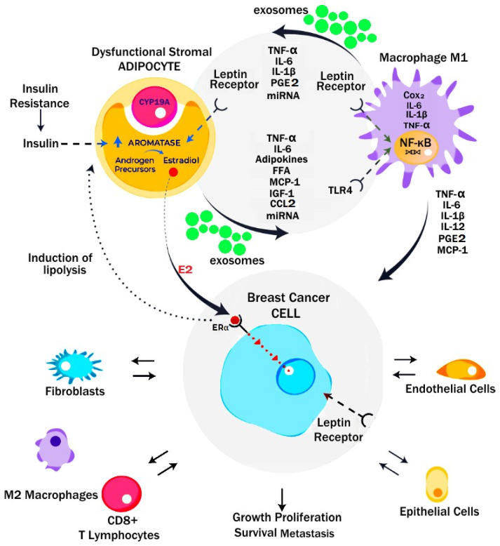
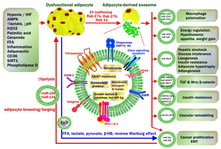
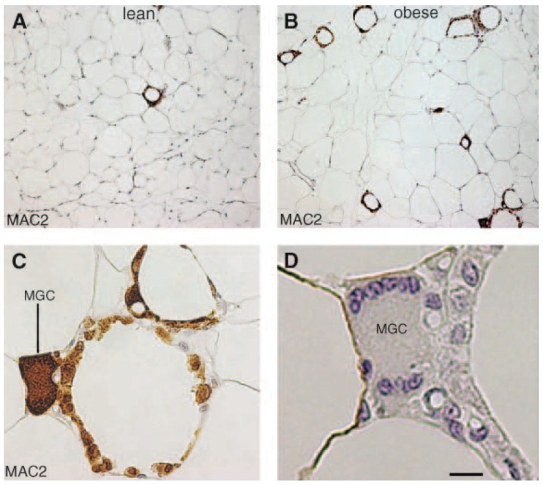
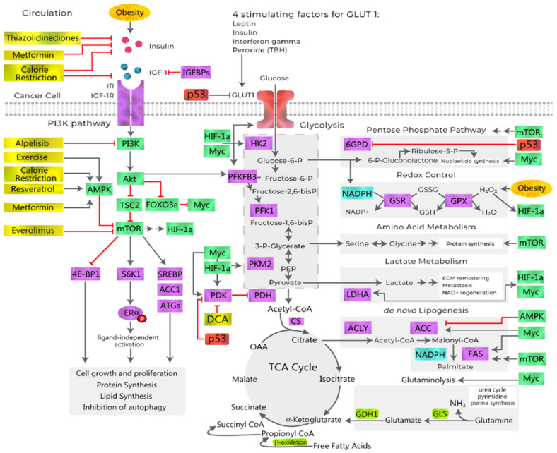
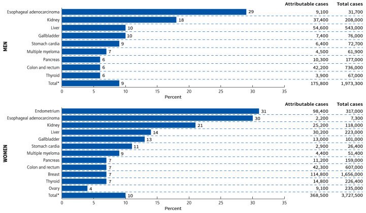
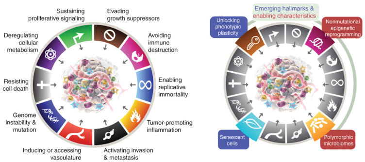
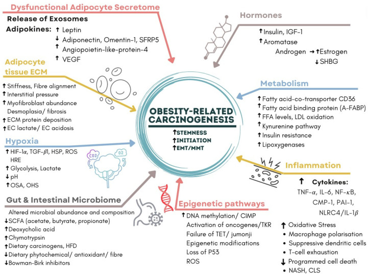
The present study aims to provide a narrative review of the molecular mechanisms of Western diet-induced obesity and obesity-related carcinogenesis. A literature search of the Cochrane Library, Embase and Pubmed databases, Google Scholar and the grey literature was conducted. Most of the molecular mechanisms that induce obesity are also involved in the twelve Hallmarks of Cancer, with the fundamental process being the consumption of a highly processed, energy-dense diet and the deposition of fat in white adipose tissue and the liver. The generation of crown-like structures, with macrophages surrounding senescent or necrotic adipocytes or hepatocytes, leads to a perpetual state of chronic inflammation, oxidative stress, hyperinsulinaemia, aromatase activity, activation of oncogenic pathways and loss of normal homeostasis. Metabolic reprogramming, epithelial mesenchymal transition, HIF-1α signalling, angiogenesis and loss of normal host immune-surveillance are particularly important. Obesity-associated carcinogenesis is closely related to metabolic syndrome, hypoxia, visceral adipose tissue dysfunction, oestrogen synthesis and detrimental cytokine, adipokine and exosomal miRNA release. This is particularly important in the pathogenesis of oestrogen-sensitive cancers, including breast, endometrial, ovarian and thyroid cancer, but also 'non-hormonal' obesity-associated cancers such as cardio-oesophageal, colorectal, renal, pancreatic, gallbladder and hepatocellular adenocarcinoma. Effective weight loss interventions may improve the future incidence of overall and obesity-associated cancer.

Keywords: GLP-1; HIF-1α; NASH; bariatric surgery; breast cancer; colorectal carcinoma; crown-like structure (CLS); cytokine; exosome; hypoxia; leptin; macrophage polarization; metabolic syndrome; obesity; senescence.


Conflict of interest statement

R.B.W. has received funding for research, education and attendance at scientific meetings from MSD, Fisher and Paykel, Ethicon and Medtronic.

Figures

Figure 5
Figure 5
Pathways of obesity-related breast cancer due to dysfunctional adipocyte secretome, with activation of PI3K/AKT/mTOR, JAK/STAT, NF-κB, RAS/RAF/MAPK and HIF/VEGF/PAI pathways. The central role of crosstalk between ER-α, IGF-1R, insulin, leptin and human EGFR in activating breast cancer cellular proliferation, survival, invasion and angiogenesis is emphasized. (Reproduced with permission from [73]). Copyright © 2023 Elsevier.
Figure 6
Figure 6
Microenvironment of dysfunctional adipocytes and breast cancer in obesity. Dysfunctional adipocytes release adipokines, cytokines and exosomes which drive a pro-inflammatory M1 phenotype in macrophages. Generation of PGE2 by tissue cyclo-oxygenase, leptin, IL-6, TNF-α and NF-κB, and loss of p53 all contribute to activation of CYP19A and production of aromatase. This leads to release of 17-β-oestradiol and activation of ER-α receptors on ER-positive breast cancer and GPER in triple-negative breast cancer. Crosstalk between cancer cells and stromal adipocytes and macrophages promotes tumour growth, survival and proliferation. In addition, recruitment of other stromal cell elements by cancer cells enables angiogenesis, evasion of T-cell immunosurveillance and chemotherapy resistance in the tumour microenvironment. (Reprinted/adapted with permission from [73]). Copyright © 2023 Elsevier.
Figure 7
Figure 7
Environmental and metabolic factors that drive dysfunctional adipocyte release of exosomes in the progression of metabolic syndrome and obesity-related carcinogenesis. This is closely related to a hypoxic, acidic, inflammatory microenvironment with high levels of oxidative stress and free radical generation. Exosome cargo includes miRNA, endosomal sorting complexes required for transport (ESCRT), cytoskeleton proteins, growth factors (TGF-1β, TNF-α, TRAIL) and receptors (EGFR), lipids, secretory signal peptides and enzymes for lipogenesis or lipolysis. During obesity, exosomal miRNA from white adipocyte tissue affect local WAT as well as cells in distant organs, including stimulation of M1 macrophage polarization, hypothalamic energy intake, WAT lipogenesis, hepatic steatosis, insulin resistance, vascular proliferation and carcinogenesis. Crosstalk from cancer cells also involves transfer of miRNA back to cancer-associated adipocytes (CAA). This stimulates adipocyte glycolysis and lipolysis and transfer of high-energy substrates including free fatty acids (FFA), lactate, pyruvate, beta-hydroxy butyrate (β-HB) to cancer cells to continue mitochondrial production of ATP (Reverse Warburg effect). Fatty acid oxidase (FAO) enzymes to assist cancer cells to utilize FFA are transferred from CAA to cancer cells via exosomes. Adapted with permission from [78]. Copyright © 2023 Springer Nature.
Figure 8
Figure 8
White adipose tissue (WAT) macrophages localize to crown-like structures (CLSs) around individual adipocytes, which increase in frequency with obesity. Light microscopy of visceral WAT of lean (A) and obese db/db (B) mouse showing MAC-2 immuno-reactive macrophages (brown colour) aggregated to form rare (A; lean) or numerous (B; obese) CLS among unilocular adipocytes. Note that almost all MAC-2 immunoreactive macrophages are organized to form CLS. (C) Enlargement of the bottom right corner of B showing that almost all mononuclear cells in CLSs are MAC-2 immunoreactive (i.e., activated macrophages). Note the multinucleate giant cell (MGC), which stains intensely for MAC-2. (D) Serial section consecutive to that shown in C confirming the presence of multiple nuclei (blue) in the MGC. Bar 100 mm for A, B, 28 mm for C, and 10 mm for D. Reproduced with permission from [112]. Copyright © 2023 Elsevier.
Figure 9
Figure 9
Obesity and activation of PI3K/AKT/mTORC1 pathways by insulin and IGF-1 in cancer cells. GLUT-1 is the predominant glucose transporter in cancers. Leptin, insulin, interferon-γ and reactive oxygen species from peroxides all stimulate GLUT-1 transport of glucose, which is fed into the hexokinase glycolysis pathway. Anabolic pathways are required for protein, nucleotide and lipid synthesis, cell growth and proliferation, and are driven by the downstream targets of mTORC1 and intracellular effects of insulin. mTORC1 also inhibits autophagy by phosphorylating proteins involved in the formation of autophagosomes (ATGs). The pentose phosphate pathway (PPP) which produces nucleotides for RNA via Ribulose-5-P, and NADPH for redox control; amino acid metabolism; lactate metabolism; and lipogenesis are controlled by AMPK, HIF-1α, mTOR and Myc. TP53 inhibits glucose flux down the PPP by regulating the dimerization of Glucose 6-phosphate dehydrogenase (G-6-PD) and inhibiting GLUT-1 and PDK. HIF-1α and c-Myc increase the activity of glucose transporters (GLUT-1), glycolytic enzymes (hexokinase-2 (HK2), phosphofructokinase (PFK1), triosephosphate isomerase (TPI1), phosphoglycerate kinase-1 (PGK-1), pyruvate kinase (PKM2) and pyruvate dehydrogenase kinase (PDHK/PDK) which inhibit PDH, driving lactate production from pyruvate via lactate dehydrogenase A (LDHA) and regeneration of NAD+. Lactate and peroxides both can stabilize HIF-1α, leading to higher activity of LDHA. Medical interventions aid in the inhibition of insulin, IGF-1 and PI3K/AKT/mTORC1/S6K1/ER-α signalling, (shown in gold). Ketone bodies, lactate and glutamine are produced by CAFs and utilized by cancer cells to maintain oxidative phosphorylation for anaplerosis. Glutamine is converted to glutamate and ammonia by GLS and then to alpha ketoglutarate by GDH-1 to enter the TCA cycle. Cytoplasmic glutamate is converted to glutathione (GSH) by glutathione synthase and used for antioxidant scavenging of ROS. Propionate is converted to succinyl-Co-A and then to succinate and fed into the TCA cycle. IGFBPs, IGF-binding proteins; IR, insulin receptor; IGF-1R, IGF-1 receptor; AMPK, AMP-activated protein kinase; TSC2, tuberous sclerosis 2; FOXO3a, forkhead box O3a; Myc, c-Myc; mTOR, mammalian target of rapamycin; HIF-1α, hypoxia-inducible factor 1-alpha; 4E-BP1, eukaryotic translation initiation factor 4E-binding protein 1; S6K1, ribosomal protein S6 kinase beta-1; SREBP, sterol regulatory element-binding protein; PFKFB3, 6-phosphofructo-2-kinase/fructose-2,6-bisphosphatase3; PEP, phosphoenolpyruvate; CS, citrate synthase; OAA, oxaloacetate; G6PD, glucose-6-phosphate dehydrogenase; GSR, glutathione reductase; GSX, glutathione peroxidase; ECM, extracellular matrix; ACLY, ATP citrate lyase; ACC, acetyl-CoA carboxylase; FAS, fatty acid synthase; DCA, dichloroacetate. (Adapted from [125]). Copyright © 2023 Doerstling, O’Flanagan and Hursting. This is an open-access article distributed under the terms of the Creative Commons Attribution License (CC BY).
Figure 1
Figure 1
Regional contribution to global obesity burden among adults by sex in 1975 and 2016. Obesity was defined as a body mass index ≥30 kg/m2. Reprinted with permission from [3]. Copyright © 2023 Wiley.
Figure 2
Figure 2
Proportions and numbers of cancer cases attributable to excess body weight (Body Mass Index ≥ 25 kg/m2) by sex and cancer type in 2012. * Total percentage is calculated among the excess body weight-related cancers listed in the figure rather than among all cancers. Figure reprinted with permission from [3]. Copyright © 2023 Wiley. Data source re-use with permission from Pearson-Stuttard, J.; Zhou, B.; Kontis, V.; Bentham, J.; Gunter, M.J.; Ezzati, M. Worldwide burden of cancer attributable to diabetes and high body-mass index: A comparative risk assessment. Lancet Diabetes Endocrinol. 2018, 6, e6–e15.8. Copyright © 2023 Elsevier.
Figure 3
Figure 3
Hallmarks of cancer and New Dimensions. Ten hallmarks of cancer with 2 emerging hallmarks and 2 enabling characteristics. Obesity and carcinogenesis share many of the same molecular mechanisms. Reprinted with permission from [26]. Copyright © 2023 American Association for Cancer Research.
Figure 4
Figure 4
Multifactorial nature of obesity-related carcinogenesis. This involves 7 categories, which are closely related to the 14 Hallmarks of Cancer and enabling characteristics. The acronyms include: A-FABP, adipose fatty acid binding protein; CD36, Cluster of differentiation; CIMP, CpG dinucleotide island methylator phenotype; CLS, crown-like structure; CMP-1, chemoattractant monocyte protein-1; ECM, extracellular matrix; EMT, epithelial-mesenchymal transition; HFD, high-fat diet; HIF-1α, hypoxia inducible factor; HSP, heat shock protein; IGF-1, insulin-like growth factor-1; IL-1, interleukin-1; IL-6, interleukin-6; LDL, low density lipoprotein; MMT, mesothelial-mesenchymal transition; NASH, non-alcoholic steatohepatitis; NF-κB, nuclear factor kappa light chain enhancer of activated B cells; NLRC4, nucleotide-binding domain and leucine-rich repeat receptor family CARD domain containing 4; OHS, obesity hypoventilation syndrome; OSA, obstructive sleep apnoea; PAI-1, Plasminogen activator inhibitor-1, ROS, reactive oxygen species; SCFA, short chain fatty acids; SFRP5, secreted frizzled-related protein 5; SHBG, sex hormone binding globulin; TGF-β1, transforming growth factor-β1; TET, ten-eleven translocation methylcytosine dioxygenase; TKR, tyrosine kinase receptor; TNF-α, tumour necrosis factor-α; VEGF, vascular endothelial growth factor. Adapted from [18]. Cellular mechanisms linking cancers to obesity by Liu et al. [18] is licensed under a Creative Commons Attribution 4.0 International License.
Figure 10
Figure 10
Metabolic crosstalk between cancer cells and CAF/CAAs: the reverse Warburg Effect. CAF and CAAs fuel the cancer cell TCA cycle by directly providing cancer cells with organic acids (lactate, pyruvate) and amino acids (alanine, glutamine) via increased glycolysis and monocarboxylate/amino acid transporters, or indirectly by enhancing glycolysis via cytokine release. This results in an increase in mitochondrial activity, leading to energy and biosynthetic precursor production and redox state modulation. CAFs also enhance mitochondrial activity in cancer cells via direct transfer of intact mitochondria. In turn, cancer cells induce aerobic glycolysis and mitochondrial dysfunction, mitophagy and ROS production in CAFs by TGF-β, integrin-β4 and exosomal miR-105 transfer, amplifying their mutual support. EMT: Epithelial Mesenchymal Transition, ETC: Electron Transport Chain, GSH: reduced glutathione, mtDNA: Mitochondrial DNA, MCT: MonoCarboxylate Transporter, ROS: Reactive Oxygen Species, TCA cycle: TriCarboxylic Acid cycle. Reproduced with permission from [133]. Attribution 4.0 International (CC BY 4.0).
Figure 11
Figure 11
The different regulation of ketone bodies between normal and cancer cells under low-carbohydrate conditions. Normal cells can catabolize ketone bodies into acetyl-CoA, which enters the TCA cycle to produce energy and improve cell viability under low-carbohydrate condition. However, excessive electrons produced by NADH/FADH2 in enhanced TCA cycle are transported to the mitochondrial respiratory chain in cancer cells, which generate ROS. In addition, the antioxidant system (PPP) pathway is inhibited under low-carbohydrate conditions, which results in excessive ROS production and leads to oxidative stress damage. Reprinted with permission from [217]. Copyright © 2023 Elsevier.

References

    1. Australian Institute of Health and Welfare Cancer Data in Australia. Cat. No. CAN 122. [(accessed on 29 November 2022)]; Available online: https://www.aihw.gov.au/reports/cancer/cancer-data-in-australia/data.
    1. Sung H., Ferlay J., Siegel R.L., Laversanne M., Soerjomataram I., Jemal A., Bray F. Global Cancer Statistics 2020: GLO-BOCAN Estimates of Incidence and Mortality Worldwide for 36 Cancers in 185 Countries. CA Cancer J. Clin. 2021;71:209–249. doi: 10.3322/caac.21660. - DOI - PubMed
    1. Sung H., Siegel R.L., Torre L.A., Pearson-Stuttard J., Islami F., Fedewa S.A., Sauer A.G., Shuval K., Gapstur S.M., Jacobs E.J. Global patterns in excess body weight and the associated cancer burden. CA Cancer J. Clin. 2019;69:88–112. doi: 10.3322/caac.21499. - DOI - PubMed
    1. Murray C.J., Aravkin A.Y., Zheng P., Abbafati C., Abbas K.M., Abbasi-Kangevari M., Abd-Allah F., Abdelalim A., Abdollahi M., Abdollahpour I. Global burden of 87 risk factors in 204 countries and territories, 1990–2019: A systematic analysis for the Global Burden of Disease Study. Lancet. 2019;396:1223–1249. doi: 10.1016/S0140-6736(20)30752-2. - DOI - PMC - PubMed
    1. Friedenreich C.M., Ryder-Burbidge C., McNeil J. Physical activity, obesity and sedentary behavior in cancer etiology: Epidemio-logic evidence and biologic mechanisms. Mol. Oncol. 2021;15:790–800. doi: 10.1002/1878-0261.12772. - DOI - PMC - PubMed

LinkOut - more resources