. 2023 Sep;17(3):618-630.
doi: 10.1007/s12105-023-01562-w. Epub 2023 May 26.

Loss of Caveolin-1 Expression in Tumor Cells is Associated with Increased Aggressiveness and Cell Invasion in Oral Squamous Cell Carcinoma

Affiliations

Loss of Caveolin-1 Expression in Tumor Cells is Associated with Increased Aggressiveness and Cell Invasion in Oral Squamous Cell Carcinoma

Rebeca Barros Nascimento et al. Head Neck Pathol. 2023 Sep.

Abstract

Background: Changes in Caveolin-1 (CAV-1) expression are related to tumorigenesis. The aim of this study was to evaluate the role of CAV-1 in tumor progression in oral squamous cell carcinoma (SCC) tissue samples and the effect of CAV-1 silencing on two oral tongue SCC (OTSCC) cell lines (SCC-25, from a primary tumor, and HSC-3 from lymph node metastases).

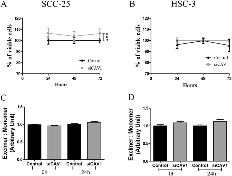
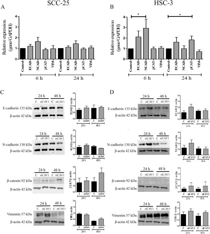
Methods: Mycroarray hybridization, mRNA expression, and immunohistochemistry were performed on OSCC tissue samples and corresponding non-tumoral margin tissues. The effects of CAV-1 silencing (siCAV-1) on cell viability, membrane fluidity, on the expression of epithelial to mesenchymal transition (EMT) markers and on cell migration and invasion capacity of OTSCC cell lines were evaluated.

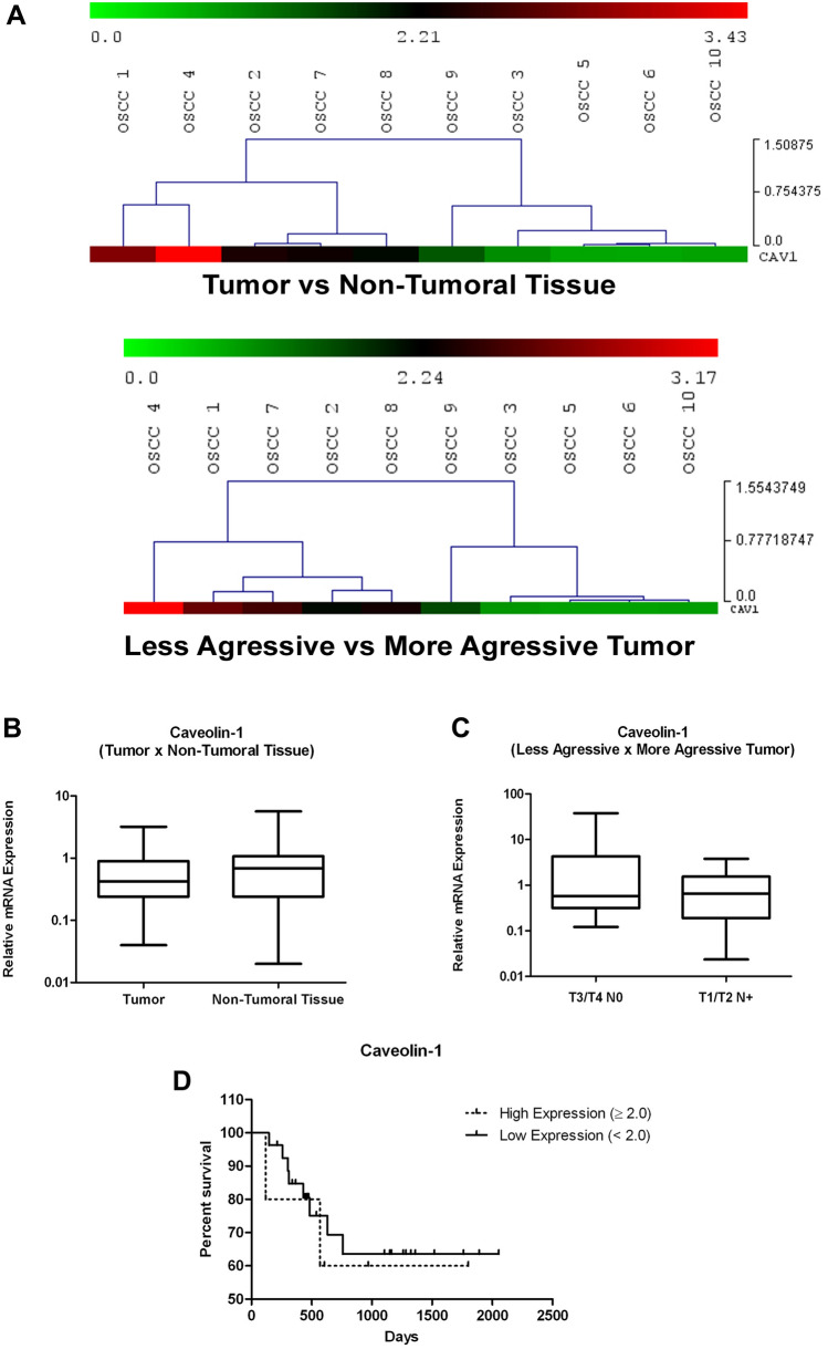
Results: Microarray showed a greater CAV-1 expression (1.77-fold) in OSCC tumors than in non-tumoral tissues and 2.0-fold more in less aggressive OSCCs. However, significant differences in CAV-1 gene expression were not seen between tumors and non-tumoral margins nor CAV-1 with any clinicopathological parameters. CAV-1 protein was localized both in carcinoma and in spindle cells of the tumor microenvironment (TME), and CAV-1 positive TME cells were associated with smaller/more aggressive tumors, independent of the carcinoma cells' expression. Silencing of CAV-1 increased cell viability only in SCC-25 cells. It also stimulated the invasion of HSC-3 cells and increased ECAD and BCAT mRNA in these cells; however, the protein levels of the EMT markers were not affected.

Conclusion: Decreased expression of CAV-1 by tumor cells in OSCC and an increase in the TME were associated with increased cell invasiveness and tumor aggressiveness.

Keywords: Caveolae; Caveolin-1; Epithelial-mesenchymal transition; Oral tongue squamous cell carcinoma; Silencing of the gene; Tumor aggressiveness.


Conflict of interest statement

The authors declare no potential conflicts of interest with respect to the research, authorship, and/or publication of this article.

Figures

Fig. 1
Fig. 1
A Hierarchical cluster diagram of CAV-1 expression in OSCC samples. Gene expression levels in non-tumoral tissues were used as the baseline. Data are visualized colorimetrically with heat plots, “red” representing elevated gene expression and “green” decreased gene expression in tumor vs non-tumoral tissue and less aggressive vs more aggressive tumors. Relative expression ratio (log10) of CAV-1 mRNA expression analysis by qRT-PCR in OSCC samples. Housekeeping gene: HPRT1. B and C Differential gene expression in tumor vs non-tumoral tissue, and less vs more aggressive tumor. D Overall survival of OSCC patients according to positive and negative CAV-1 gene expression
Fig. 2
Fig. 2
Immunoexpression of CAV-1 in OSCC and non-tumoral tissue. CAV-1 shows cytoplasmic immunoexpression in matched non-tumoral tissue in basal /suprabasal layers (A), intense membranous/cytoplasmic immunoexpression in carcinoma (B and C), and TME (C and D). Overall survival of OSCC patients according to positive and negative CAV-1 in carcinoma (E) and TME (F) immunoexpression (Ep epithelium; St: stroma; T: tumor; BV: blood vessel)
Fig. 3
Fig. 3
Effect of siCAV-1 in cell viability and membrane fluidity. Analysis of % of viable cells by Alamar Blue assay after siCAV-1 in SCC-25 cells (A) and HSC-3 cells (B). Evaluation of membrane fluidity by fluorescence assay using a Membrane Fluidity kit in SCC-25 (C) and HSC-3 (D) cells after siCAV-1. (***p < 0.001; Error bars represent the standard deviation of 3 or more experiments)
Fig. 4
Fig. 4
Effect of siCAV-1 on gene and protein expression. Analysis of gene expression of EMT markers by qRT-PCR in SCC-25 cells (A) and HSC-3 cells (B). Western Blotting experiments for evaluation of protein expression of EMT markers in SCC-25 cells (C) and HSC-3 cells (D). (*p < 0.05; Error bars represent the standard deviation of 3 or more different experiments)
Fig. 5
Fig. 5
Effect of siCAV-1 on cell migration and invasion. Migration assay was evaluated by scratch wound healing assay on Myogel coating in both SCC-25 (A and B) and HSC-3 (C and D) cells after 48 h siCAV-1. A wound healing assay was performed in Myogel-fibrin for evaluating the effect of silencing of CAV-1 on the invasion capacity of SCC-25 (E and F) and HSC-3 (G and H) cells (*p < 0.05, **p < 0.01, ***p < 0.001; Error bars represent the standard deviation of 3 or more different experiments)
Fig. 6
Fig. 6
Summary of results. Normal cells present cholesterol-rich membranes with the presence of caveolar structures containing Caveolin-1 (CAV-1) protein. An increase in CAV-1 expression was observed in tumors when compared to non-tumor margin tissues and in more aggressive tumors when compared to less aggressive tumors. This increase in CAV-1 expression was greater in tumor microenvironment cells, such as cancer-associated fibroblasts. In addition, the silencing of CAV-1 in OSCC cells induced an increase in the cell viability of non-metastatic SCC-25 cells but increased the invasive capacity of metastatic HSC-3 cells, showing that the effect of CAV-1 expression varies in different cellular profiles of the same tumor type

References

    1. Bray F, Ferlay J, Soerjomataram I, et al. Global cancer statistics 2018: globocan estimates of incidence and mortality worldwide for 36 cancers in 185 countries. CA Cancer J Clin. 2018;68:394–424. doi: 10.3322/caac.21492. - DOI - PubMed
    1. Flavahan WA, Gaskell E, Bernstein BE. Epigenetic plasticity and the hallmarks of cancer. Science. 2017 doi: 10.1126/science.aal2380. - DOI - PMC - PubMed
    1. Farah CS. Molecular landscape of head and neck cancer and implications for therapy. Ann Transl Med. 2021 doi: 10.21037/atm-20-6264. - DOI - PMC - PubMed
    1. Campos A, Burgos-Ravanal R, González MF, et al. Cell intrinsic and extrinsic mechanisms of caveolin-1-enhanced metastasis. Biomolecules. 2019 doi: 10.3390/biom9080314. - DOI - PMC - PubMed
    1. Routray S. Caveolin-1 in oral squamous cell carcinoma microenvironment: an overview. Tumor Biol. 2014;35:9487–9495. doi: 10.1007/s13277-014-2482-z. - DOI - PubMed