. 2023 Apr 29;26(5):106766.
doi: 10.1016/j.isci.2023.106766. eCollection 2023 May 19.

A cell transmembrane peptide chimeric M(27-39)-HTPP targeted therapy for hepatocellular carcinoma

Affiliations

A cell transmembrane peptide chimeric M(27-39)-HTPP targeted therapy for hepatocellular carcinoma

Jibin Wu et al. iScience. .

Abstract

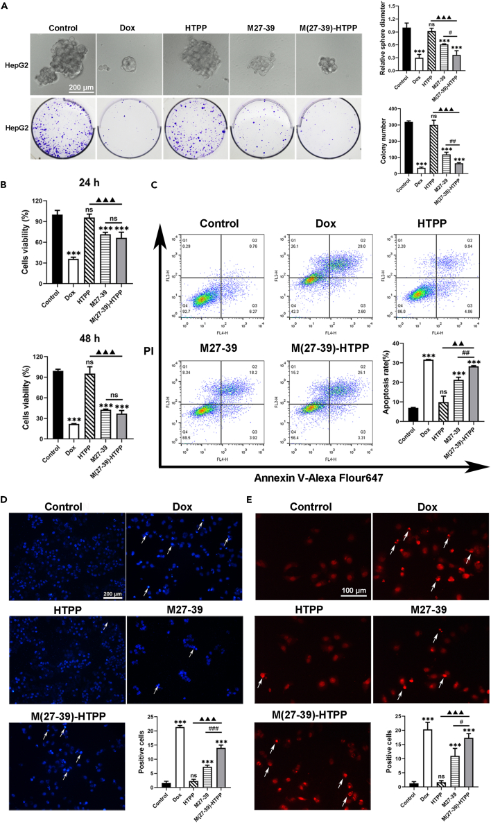
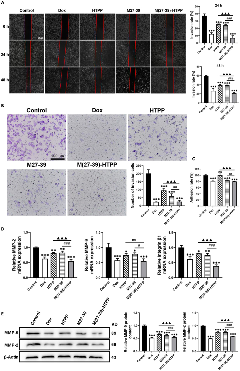
Hepatocellular carcinoma (HCC) is a prevalent malignant tumor, with a growing incidence and death rate worldwide. The aims and challenges of treating HCC include targeting the tumor, entering the tumor tissue, inhibiting the spread and growth of tumor cells. M27-39 is a small peptide isolated from the antimicrobial peptide Musca domestica cecropin (MDC), whereas HTPP is a liver-targeting, cell-penetrating peptide obtained from the circumsporozoite protein (CSP) of Plasmodium parasites. In this study, M27-39 was modified by HTPP to form M(27-39)-HTPP, which targeted tumor penetration to treat HCC. Here, we revealed that M(27-39)-HTPP had a good ability to target and penetrate the tumor, effectively limit the proliferation, migration, and invasion, and induce the apoptosis in HCC. Notably, M(27-39)-HTPP demonstrated good biosecurity when administered at therapeutic doses. Accordingly, M(27-39)-HTPP could be used as a new, safe, and efficient therapeutic peptide for HCC.

Keywords: Cancer; Chemistry; Oncology.


Conflict of interest statement

The patent in question is held by X.L./Guangdong Pharmaceutical University, ZhaoXia Chen, S.G., Lun Zhang, Along Liu, Yanan Tang, Jie Wang, Wenbin Liu, X.J., Jiayong Zhu. (Patent No. ZL201810196235.0). The authors declare no conflict of interest.

Figures

None
Graphical abstract
Figure 1
Figure 1
Evaluation of in vitro targeted uptake of M(27–39)-HTPP (A) Localization of M(27–39)-HTPP in HepG2 cells. (B) Assessment of M(27–39)-HTPP and M27-39 uptake in HepG2 cells (Microscopes). (C) Assessment of M(27–39)-HTPP and M27-39 uptake in HepG2 cells (Flow cytometry). (D) Assessment of M(27–39)-HTPP uptake HepG2, L02, MCF7, HCT116 and A549 cells (Microscopes). (E) Assessment of M(27–39)-HTPP uptake HepG2, L02, MCF7, HCT116 and A549 cells (Flow cytometry). (F) Comparison of penetration of M(27–39)-HTPP and M27-39 into HepG2 tumor spheres. (G) M(27–39)-HTPP colocalizes with GPC3. (H) GPC3 expression after transfection with GPC3 siRNA. (I) HepG2 cells incubated with FITC-HTPP by pre-treating with siRNA interference (Flow cytometry). (J) HepG2 cells incubated with FITC-M(27–39)-HTPP by pre-treating with siRNA interference (Flow cytometry).∗p< 0.05; ∗∗p< 0.01; ∗∗∗p< 0.001. as compared with the negative control group. #p< 0.05; ##p< 0.01; ###p< 0.001. p< 0.05; ▲▲p< 0.01; ▲▲▲p< 0.001. p< 0.05; ★★p< 0.01; ★★★p< 0.001. N.S., not significant. Data are presented as the mean ± SD.
Figure 2
Figure 2
In vivo targeted penetration of M(27–39)-HTPP (A) Imaging of small animals in vivo. (B–D) Comparison of the fluorescence intensity of each organ in M(27–39)-HTPP group, and comparison of the fluorescence intensity of liver between M(27–39)-HTPP and M27-39 group. (E and F) Localization of M(27–39)-HTPP and M27-39 in mouse liver. ∗p< 0.05; ∗∗p< 0.01; ∗∗∗p< 0.001. as compared with the negative control group. N.S., not significant. Data are presented as the mean ± SD.
Figure 3
Figure 3
In vitro inhibition of HepG2 cells by M(27–39)-HTPP (A) Cell viability of the HepG2 cells treated with various drugs. (B) Inhibition of tumor globule and colony formation in HepG2 by M(27–39)-HTPP. (C) Apoptosis of the HepG2 cells induced by M(27–39)-HTPP, statistical analysis of apoptosis rate. (D and E) HepG2 cells were stained for apoptotic cells, using and hoechst33324 and TUNEL assay staining; arrows, representative apoptotic cells with hoechst33324-positive and TUNEL-positive. ∗p< 0.05; ∗∗p< 0.01; ∗∗∗p< 0.001. As compared with the negative control group. #p< 0.05; ##p< 0.01; ###p< 0.001. p< 0.05; ▲▲p< 0.01; ▲▲▲p< 0.001. N.S., not significant. Data are presented as the mean ± SD.
Figure 4
Figure 4
Effect of M(27–39)-HTPP on the expression of apoptosis-related genes and proteins in HepG2 cells (A) Relative mRNA expression of P53, Bcl-2, Bax, Caspase-9, Caspase-3 and PARP in HepG2 cells. (B) Western blot and relative protein expression of P53, Bcl-2, Bax, Cyt-C, Cleaved Caspase-9, Cleaved Caspase-3 and Cleaved PARP in HepG2 cells. ∗p< 0.05; ∗∗p< 0.01; ∗∗∗p< 0.001. As compared with the negative control group. #p< 0.05; ##p< 0.01; ###p< 0.001. p< 0.05; ▲▲p< 0.01; ▲▲▲p< 0.001. N.S., not significant. Data are presented as the mean ± SD.
Figure 5
Figure 5
Effects of M(27–39)-HTPP on migration, invasion and adhesion of HepG2 cells (A) Wound healing assay measured the migration of the HepG2 cells. (B) Transwell assay evaluated the invasion of the HepG2 cells. (C) Adhesion ability of HepG2 cells. (D) Relative mRNA expression of MMP-2, MMP-9 and Integrin β1 in HepG2 cells. (E) Western blot and relative protein expression of MMP-2 and MMP-9 in HepG2 cells.∗p< 0.05; ∗∗p< 0.01; ∗∗∗p< 0.001. as compared with the negative control group. #p< 0.05; ##p< 0.01; ###p< 0.001. p< 0.05; ▲▲p< 0.01; ▲▲▲p< 0.001. N.S., not significant. Data are presented as the mean ± SD.
Figure 6
Figure 6
Anti-tumor effect of M(27–39)-HTPP in vivo (A and B) Growth status and body weight of nude mice (n = 6). (C) Hepatoma size in nude mice (n = 6). ∗p< 0.05; ∗∗p< 0.01; ∗∗∗p< 0.001. as compared with the negative control group. #p< 0.05; ##p< 0.01; ###p< 0.001. p< 0.05; ▲▲p< 0.01; ▲▲▲p< 0.001. N.S., not significant. Data are presented as the mean ± SD.

Similar articles

Cited by

References

    1. Sung H., Ferlay J., Siegel R.L., Laversanne M., Soerjomataram I., Jemal A., Bray F. Global cancer statistics 2020: GLOBOCAN estimates of incidence and mortality worldwide for 36 cancers in 185 countries. CA. Cancer J. Clin. 2021;71:209–249. doi: 10.3322/caac.21660. - DOI - PubMed
    1. Li G., Zhao L. Sorafenib-loaded hydroxyethyl starch-TG100-115 micelles for the treatment of liver cancer based on synergistic treatment. Drug Deliv. 2019;26:756–764. doi: 10.1080/10717544.2019.1642418. - DOI - PMC - PubMed
    1. Wang Y., Chen X., He D., Zhou Y., Qin L. Surface-modified nanoerythrocyte loading DOX for targeted liver cancer chemotherapy. Mol. Pharm. 2018;15:5728–5740. doi: 10.1021/acs.molpharmaceut.8b00881. - DOI - PubMed
    1. Xu J., Cheng X., Tan L., Fu C., Ahmed M., Tian J., Dou J., Zhou Q., Ren X., Wu Q., et al. Microwave responsive nanoplatform via P-selectin mediated drug delivery for treatment of hepatocellular carcinoma with distant metastasis. Nano Lett. 2019;19:2914–2927. doi: 10.1021/acs.nanolett.8b05202. - DOI - PubMed
    1. Gong X., Zheng Y., He G., Chen K., Zeng X., Chen Z. Multifunctional nanoplatform based on star-shaped copolymer for liver cancer targeting therapy. Drug Deliv. 2019;26:595–603. doi: 10.1080/10717544.2019.1625467. - DOI - PMC - PubMed

LinkOut - more resources