

From Karl Wurm and Guy Scadding's staging to 18F-FDG PET/CT scan phenotyping and far beyond: perspective in the evading history of phenotyping in sarcoidosis
- PMID: 37234239
- PMCID: PMC10206027
- DOI: 10.3389/fmed.2023.1174518
From Karl Wurm and Guy Scadding's staging to 18F-FDG PET/CT scan phenotyping and far beyond: perspective in the evading history of phenotyping in sarcoidosis
Abstract
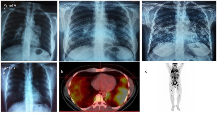
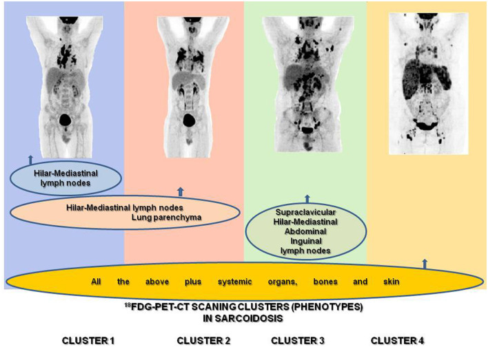
Sarcoidosis is an inflammatory granulomatous disease of unknown etiology involving any organ or tissue along with any combination of active sites, even the most silent ones clinically. The unpredictable nature of the sites involved in sarcoidosis dictates the highly variable natural history of the disease and the necessity to cluster cases at diagnosis based on clinical and/or imaging common characteristics in an attempt to classify patients based on their more homogeneous phenotypes, possibly with similar clinical behavior, prognosis, outcome, and therefore with therapeutic requirements. In the course of the disease's history, this attempt relates to the availability of a means of detection of the sites involved, from the Karl Wurm and Guy Scadding's chest x-ray staging through the ACCESS, the WASOG Sarcoidosis Organ Assessment Instruments, and the GenPhenReSa study to the 18F-FDG PET/CT scan phenotyping and far beyond to new technologies and/or the current "omics." The hybrid molecular imaging of the 18F-FDG PET/CT scan, by unveiling the glucose metabolism of inflammatory cells, can identify high sensitivity inflammatory active granulomas, the hallmark of sarcoidosis-even in clinically and physiologically silent sites-and, as recently shown, is successful in identifying an unexpected ordered stratification into four phenotypes: (I) hilar-mediastinal nodal, (II) lungs and hilar-mediastinal nodal, (III) an extended nodal supraclavicular, thoracic, abdominal, inguinal, and (IV) all the above in addition to systemic organs and tissues, which is therefore the ideal phenotyping instrument. During the "omics era," studies could provide significant, distinct, and exclusive insights into sarcoidosis phenotypes linking clinical, laboratory, imaging, and histologic characteristics with molecular signatures. In this context, the personalization of treatment for sarcoidosis patients might have reached its goal.
Keywords: 18F-FDG PET/CT scan phenotyping; chest roentgenogram staging; omics; personalized treatment; sarcoidosis.
Copyright © 2023 Papiris, Kolilekas, Rivera, Spanos, Li, Gokulnath, Chatterjee, Georgakopoulos, Kallieri, Papaioannou, Raptakis, Apollonatou, Antonogiannaki, Gialafos, Chatziioannou, Grunewald and Manali.
Conflict of interest statement
The authors declare that the research was conducted in the absence of any commercial or financial relationships that could be construed as a potential conflict of interest.
Figures
References
LinkOut - more resources
Full Text Sources