. 2023 May 2:8:76.
doi: 10.12688/wellcomeopenres.18937.2. eCollection 2023.

Structure of the transmembrane protein 2 (TMEM2) ectodomain and its apparent lack of hyaluronidase activity

Affiliations

Structure of the transmembrane protein 2 (TMEM2) ectodomain and its apparent lack of hyaluronidase activity

Muyuan Niu et al. Wellcome Open Res. .

Abstract

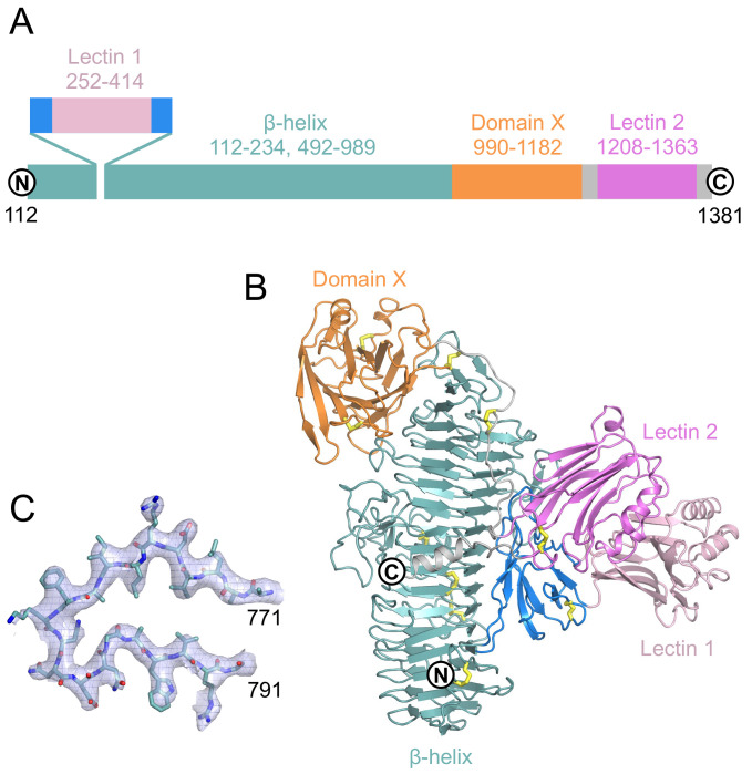
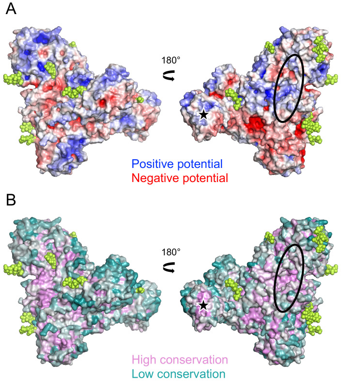
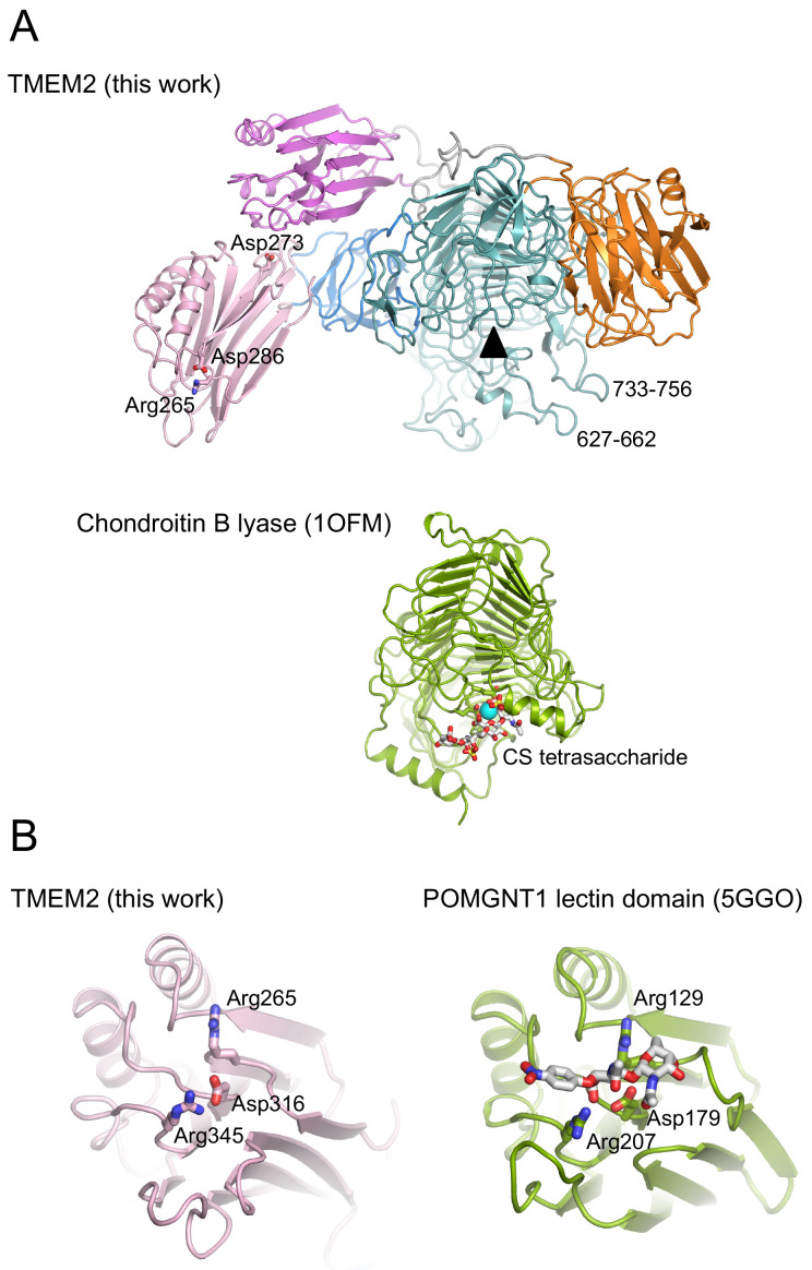
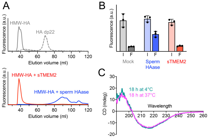
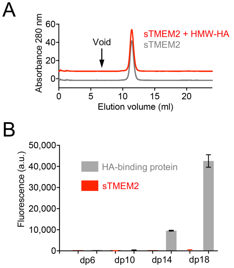
Background: Hyaluronic acid (HA) is a major polysaccharide component of the extracellular matrix. HA has essential functions in tissue architecture and the regulation of cell behaviour. HA turnover needs to be finely balanced. Increased HA degradation is associated with cancer, inflammation, and other pathological situations. Transmembrane protein 2 (TMEM2) is a cell surface protein that has been reported to degrade HA into ~5 kDa fragments and play an essential role in systemic HA turnover. Methods: We produced the soluble TMEM2 ectodomain (residues 106-1383; sTMEM2) in human embryonic kidney cells (HEK293) and determined its structure using X-ray crystallography. We tested sTMEM2 hyaluronidase activity using fluorescently labelled HA and size fractionation of reaction products. We tested HA binding in solution and using a glycan microarray. Results: Our crystal structure of sTMEM2 confirms a remarkably accurate prediction by AlphaFold. sTMEM2 contains a parallel β-helix typical of other polysaccharide-degrading enzymes, but an active site cannot be assigned with confidence. A lectin-like domain is inserted into the β-helix and predicted to be functional in carbohydrate binding. A second lectin-like domain at the C-terminus is unlikely to bind carbohydrates. We did not observe HA binding in two assay formats, suggesting a modest affinity at best. Unexpectedly, we were unable to observe any HA degradation by sTMEM2. Our negative results set an upper limit for k cat of approximately 10 -5 min -1. Conclusions: Although sTMEM2 contains domain types consistent with its suggested role in TMEM2 degradation, its hyaluronidase activity was undetectable. HA degradation by TMEM2 may require additional proteins and/or localisation at the cell surface.

Keywords: Glycosaminoglycan; X-ray crystallography; hyaluronidase; lectin; parallel β-helix.


Conflict of interest statement

No competing interests were disclosed.

Figures

Figure 1.
Figure 1.. Crystal structure of soluble Transmembrane protein 2 (sTMEM2).
( A) Schematic representation of the sTMEM2 domain structure. Sequence numbers refer to full-length TMEM2. ( B) Cartoon drawing of the sTMEM2 structure using the same colour code as in A. Disulfide bonds are indicated by yellow sticks. N-linked glycans have been omitted for clarity. ( C) Electron density map for residues 771-791. Shown is the final 2 F obs- F calc map contoured at 1 σ level. This figure was produced using PyMOL version 2.5.2. UCSF Chimera is a free alternative to PYMOL.
Figure 2.
Figure 2.. Surface properties of soluble Transmembrane protein 2 (sTMEM2).
( A) Electrostatic surface potential calculated using the APBS function of PyMOL (version 2.4.1). UCSF Chimera is a free alternative to PYMOL. Positive and negative potential is shown in blue and red, respectively. Glycans are shown in lime green. ( B) Sequence conservation calculated using the Consurf server . High and low sequence conservation is shown in magenta and teal, respectively. Glycans are shown in lime green. In A and B, the views on the left are the same as in Figure 1B. The black oval indicates the general location of active sites in related β-helix enzymes (see text). The black star indicates the tip of lectin-like domain 1.
Figure 3.
Figure 3.. Comparison of soluble Transmembrane protein 2 (sTMEM2) to other proteins.
( A) Chondroitin lyase ( PDB 1OFM) was superimposed onto the β-helix of sTMEM2 with a r.m.s. deviation of 3.3 Å for 325 Cα atoms. Chondroitin lyase contains a Ca 2+ ion in the active site; the equivalent position in sTMEM2 is indicated by the black triangle. Three residues in the lectin-like domain 1 whose mutation has been reported to reduce TMEM2 activity are shown in atomic detail. ( B) The N-terminal lectin domain of POMGNT1 ( PDB 5GGO) was superimposed onto the lectin-like domain 1 of sTMEM2 with a r.m.s. deviation of 2.3 Å for 153 Cα atoms. The disaccharide bound to POMGNT1 is shown in atomic detail, as are selected residues that are conserved between sTMEM2 and POMGNT1. This figure was produced using PyMOL version 2.5.2. UCSF Chimera is a free alternative to PYMOL.
Figure 4.
Figure 4.. Hyaluronidase activity of soluble Transmembrane protein 2 (sTMEM2).
( A) Top, size exclusion chromatography of 100 μg/ml fluorescein-labelled high-molecular-weight Hyaluronic acid (HMW-HA) or HA 22-mer (dp22, ~5 kDa). Bottom; size exclusion chromatography of 100 μg/ml fluorescein-labelled HMW-HA incubated overnight with 100 μg/ml sTMEM2 or sperm hyaluronidase (pH 6.0, 1 mM CaCl 2). Only the sperm hyaluronidase was able to degrade HMW-HA. Shown is a representative of n = 3 experiments. ( B) Ultrafiltration of 0.1 μg/ml fluorescein-labelled HMW-HA incubated overnight with 100 μg/ml sTMEM2 and sperm hylauronidase (pH 6.0, 1 mM CaCl 2). The fluorescence was measured in the solution before and after filtration (I, input; F, filtrate). Only the sperm hyaluronidase was able to degrade HMW-HA. Data are presented as mean values ± s.e.m. for n = 3 independent experiments. ( C) Circular dichroism spectra of sTMEM2 incubated overnight at 4°C (teal) and 37°C (magenta). The minimum at 217 nm is characteristic of β-sheet structure. There is no evidence of denaturation at the higher temperature. The raw measurements are available in Underlying data.
Figure 5.
Figure 5.. Hyaluronan binding by soluble Transmembrane protein 2 (sTMEM2).
( A) Size exclusion chromatography of sTMEM2 in the absence or presence of unlabelled HMW-HA. To avoid overlap of the two traces, the red trace has been shifted by +10 mAU. No sTMEM2 protein is associated with HMW-HA eluting in the void volume of the column (arrow). ( B) Binding of sTMEM2 and the aggrecan G1-link protein complex (HA-binding protein) to HA oligomers of different length (dp, degree of polymerisation). The experiment was done using a neoglycolipid-based microarray , . Data (fluorescence intensities of HA probes arrayed at 5 fmol/spot level) are presented as mean values ± range for n = 2 duplicate spots on the array. The raw measurements are available in Underlying data.

References

    1. Kobayashi T, Chanmee T, Itano N: Hyaluronan: metabolism and function. Biomolecules. 2020;10(11):1525. 10.3390/biom10111525 - DOI - PMC - PubMed
    1. Bohaumilitzky L, Huber AK, Stork EM, et al. : A trickster in disguise: hyaluronan's ambivalent roles in the matrix. Front Oncol. 2017;7:242. 10.3389/fonc.2017.00242 - DOI - PMC - PubMed
    1. Yamaguchi Y, Yamamoto H, Tobisawa Y, et al. : TMEM2: A missing link in hyaluronan catabolism identified? Matrix Biol. 2019;78–79:139–146. 10.1016/j.matbio.2018.03.020 - DOI - PMC - PubMed
    1. Yoshida H, Nagaoka A, Kusaka-Kikushima A, et al. : KIAA1199, a deafness gene of unknown function, is a new hyaluronan binding protein involved in hyaluronan depolymerization. Proc Natl Acad Sci U S A. 2013;110(14):5612–5617. 10.1073/pnas.1215432110 - DOI - PMC - PubMed
    1. Yamamoto H, Tobisawa Y, Inubushi T, et al. : A mammalian homolog of the zebrafish transmembrane protein 2 (TMEM2) is the long-sought-after cell-surface hyaluronidase. J Biol Chem. 2017;292(18):7304–7313. 10.1074/jbc.M116.770149 - DOI - PMC - PubMed