

Case Report: Giant insulinoma, a very rare tumor causing hypoglycemia
- PMID: 37234805
- PMCID: PMC10206132
- DOI: 10.3389/fendo.2023.1125772
Case Report: Giant insulinoma, a very rare tumor causing hypoglycemia
Abstract
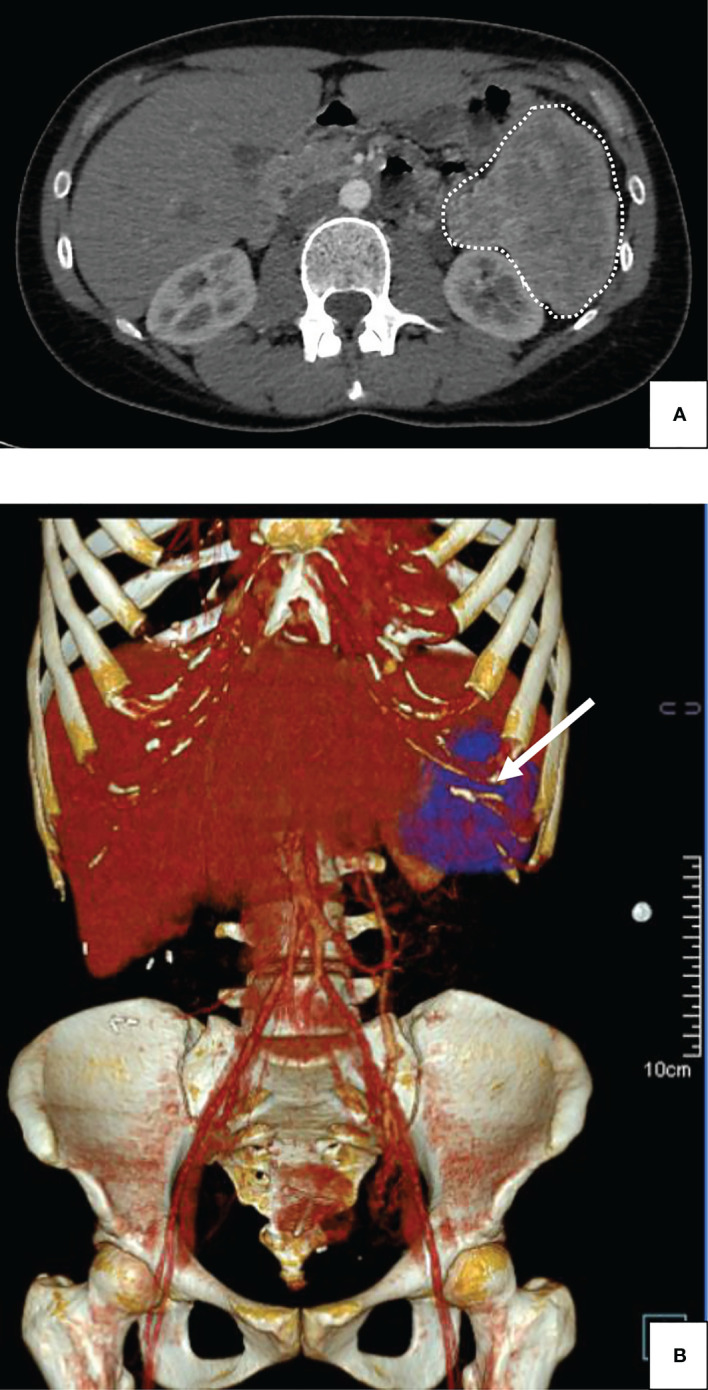
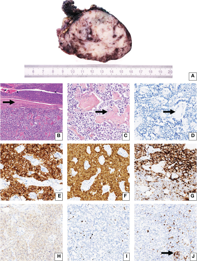
Insulinomas, with an incidence of 4 cases per million individuals per year, remain amongst the most frequent functional neuroendocrine tumors. The usual diameter of insulinomas usually remains under 3 cm of major axis. However, 44 exceptional cases of "giant insulinomas", have been reported worldwide, generally exceeding 9 cm in major axis. In this article, we report the case of a 38-year-old woman whom suffered from chronic hypoglycemia despite treatment with diazoxide. Abdominal CT-scan revealed a 88 x 73 mm mass located at the tail of the pancreas. Following surgical excision, histopathological analysis confirmed G1 neuroendocrine tumor, with focal cytoplasmic expression of insulin in tumor cells. After a 16-month follow-up period, the patient didn't address any specific complaint, and no disease recurrence and/or metastasis were observed. A 68Ga-DOTATATE-PET scan was performed 6 months after surgery, which came back normal. Genetic evaluation has not been performed in our patient. The physiopathology of giant insulinomas remain unexplained, however with possible relationship with type 1 multiple endocrine neoplasia, sporadic somatic YY1 mutations and possible transformation of bulky non-functional pancreatic neuroendocrine tumors to a functional phenotype, with slow insulin secretion. While giant insulinomas remain rare in the literature, multicentric genetic analysis of tumor samples might reveal unique features of this rare subtype of neuroendocrine pancreatic tumors. Insulinomas of large size tend to have greater malignancy and higher rates of invasiveness. Careful follow-up, especially for liver and lymph node metastases, must be performed using functional imaging techniques to avoid disease relapse.
Keywords: giant insulinoma; hypoglycemia; neuroendocrine tumors; pancreas; pathology.
Copyright © 2023 Tarris, Rouland, Guillen, Loffroy, Lariotte, Rat, Bouillet, Andrianiaina, Petit and Martin.
Conflict of interest statement
The authors declare that the research was conducted in the absence of any commercial or financial relationships that could be construed as a potential conflict of interest.
Figures
Similar articles
-
Multifocal Insulinoma as the Unique Presenting Feature of Multiple Endocrine Neoplasia Type 1 in an Adolescent.Horm Res Paediatr. 2025;98(1):75-83. doi: 10.1159/000538211. Epub 2024 Mar 5. Horm Res Paediatr. 2025. PMID: 38442699 Free PMC article.
-
Case presentation of 8-year follow up of recurrent malignant duodenal Insulinoma and lymph node metastases and literature review of malignant Insulinoma management.BMC Endocr Disord. 2022 Dec 9;22(1):310. doi: 10.1186/s12902-022-01219-9. BMC Endocr Disord. 2022. PMID: 36494838 Free PMC article. Review.
-
Radiofrequency ablation of solitary pancreatic insulinoma in a patient with episodes of severe hypoglycemia.Eur J Gastroenterol Hepatol. 2009 Sep;21(9):1097-101. doi: 10.1097/meg.0b013e328323d70e. Eur J Gastroenterol Hepatol. 2009. PMID: 19685572
-
Surgery for multiple endocrine neoplasia type 1-related insulinoma: long-term outcomes in a large international cohort.Br J Surg. 2020 Oct;107(11):1489-1499. doi: 10.1002/bjs.11632. Epub 2020 Apr 30. Br J Surg. 2020. PMID: 32352164 Free PMC article.
-
Determinants of surgical resection for pancreatic neuroendocrine tumors.J Hepatobiliary Pancreat Sci. 2015 Aug;22(8):610-7. doi: 10.1002/jhbp.224. Epub 2015 Mar 13. J Hepatobiliary Pancreat Sci. 2015. PMID: 25773163 Review.
Cited by
-
Pancreatic Neuroendocrine Tumors-Diagnostic Pitfalls of Non-Diabetic Severe Hypoglycemia: Literature Review and Case Report.Diagnostics (Basel). 2025 Jan 31;15(3):337. doi: 10.3390/diagnostics15030337. Diagnostics (Basel). 2025. PMID: 39941267 Free PMC article.
-
History of an Insidious Case of Metastatic Insulinoma.J Clin Med. 2025 Jun 6;14(12):4028. doi: 10.3390/jcm14124028. J Clin Med. 2025. PMID: 40565772 Free PMC article.
References
-
- Vaidakis D, Karoubalis J, Pappa T, Piaditis G, Zografos GN. Pancreatic insulinoma: current issues and trends. Hepatobiliary Pancreat Dis Int (2010) 9:234–41. - PubMed
Publication types
MeSH terms
LinkOut - more resources
Full Text Sources
Medical