

Bisphenol S Increases Cell Number and Stimulates Migration of Endometrial Epithelial Cells
- PMID: 37234927
- PMCID: PMC10207871
- DOI: 10.15605/jafes.037.S7
Bisphenol S Increases Cell Number and Stimulates Migration of Endometrial Epithelial Cells
Abstract
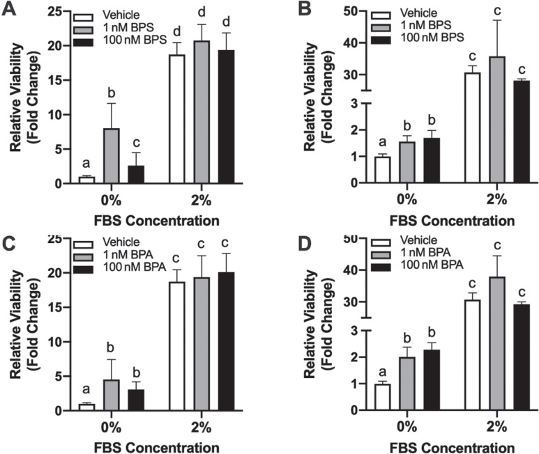
Objective: To determine whether bisphenol S (BPS), a common substitute for bisphenol A (BPA), induces cell proliferation and migration in human endometrial epithelial cells (Ishikawa) and adult mouse uterine tissues.
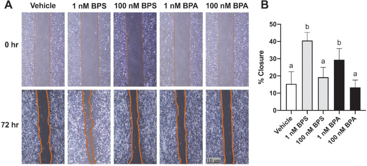
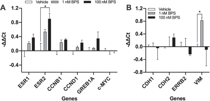
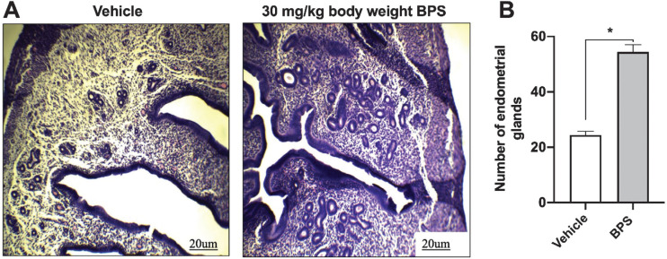
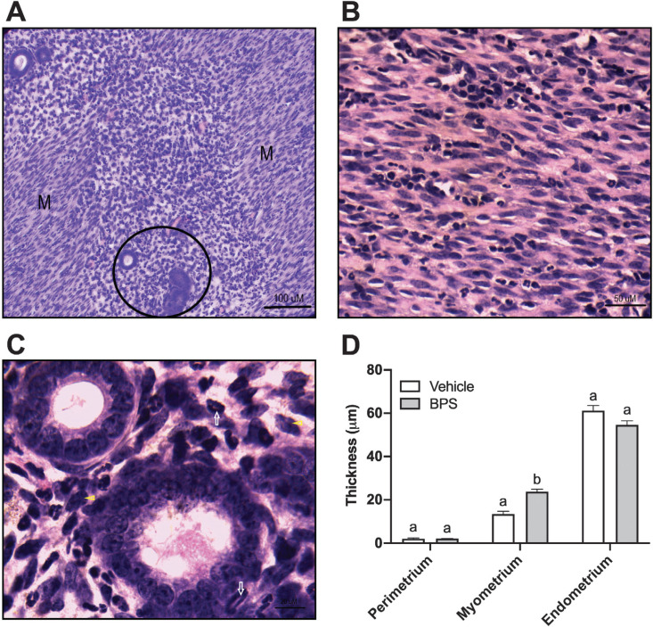
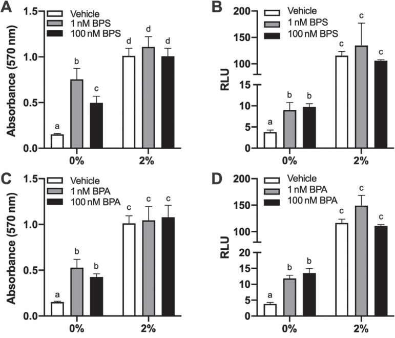
Methodology: Human endometrial Ishikawa cells were exposed to low doses of BPS (1 nM and 100 nM) for 72 hours. Cell proliferation was assessed through the viability assays MTT and CellTiter-Glo®. Wound healing assays were also used to evaluate the migration potential of the cell line. The expression of genes related to proliferation and migration was also determined. Similarly, adult mice were exposed to BPS at a dose of 30 mg/kg body weight/day for 21 days, after which, the uterus was sent for histopathologic assessment.
Results: BPS increased cell number and stimulated migration in Ishikawa cells, in association with the upregulation of estrogen receptor beta (ESR2) and vimentin (VIM). In addition, mice exposed to BPS showed a significantly higher mean number of endometrial glands within the endometrium.
Conclusion: Overall, in vitro and in vivo results obtained in this study showed that BPS could significantly promote endometrial epithelial cell proliferation and migration, a phenotype also observed with BPA exposure. Hence, the use of BPS in BPA-free products must be reassessed, as it may pose adverse reproductive health effects to humans.
Keywords: BPS; Ishikawa cells; endocrine-disrupting chemicals; hyperplasia; uterus.
© 2022 Journal of the ASEAN Federation of Endocrine Societies.
Conflict of interest statement
The authors declared no conflict of interest.
Figures
Similar articles
-
Bisphenol A Analogues Suppress Spheroid Attachment on Human Endometrial Epithelial Cells through Modulation of Steroid Hormone Receptors Signaling Pathway.Cells. 2021 Oct 26;10(11):2882. doi: 10.3390/cells10112882. Cells. 2021. PMID: 34831106 Free PMC article.
-
Environmentally relevant levels of bisphenol A affect uterine decidualization and embryo implantation through the estrogen receptor/serum and glucocorticoid-regulated kinase 1/epithelial sodium ion channel α-subunit pathway in a mouse model.Fertil Steril. 2018 Apr;109(4):735-744.e1. doi: 10.1016/j.fertnstert.2017.12.003. Epub 2018 Mar 28. Fertil Steril. 2018. PMID: 29605410
-
Developmental exposures to bisphenol S, a BPA replacement, alter estrogen-responsiveness of the female reproductive tract: a pilot study.Cogent Med. 2017;4(1):1317690. Epub 2017 Apr 26. Cogent Med. 2017. PMID: 31231671 Free PMC article.
-
Occurrence of bisphenol S in the environment and implications for human exposure: A short review.Sci Total Environ. 2018 Feb 15;615:87-98. doi: 10.1016/j.scitotenv.2017.09.194. Epub 2017 Oct 17. Sci Total Environ. 2018. PMID: 28963899 Review.
-
A new chapter in the bisphenol A story: bisphenol S and bisphenol F are not safe alternatives to this compound.Fertil Steril. 2015 Jan;103(1):11-21. doi: 10.1016/j.fertnstert.2014.11.005. Epub 2014 Dec 2. Fertil Steril. 2015. PMID: 25475787 Review.
Cited by
-
Influence of bisphenol A and its analog bisphenol S on cocaine- and amphetamine-regulated transcript peptide-positive enteric neurons in the mouse gastrointestinal tract.Front Mol Neurosci. 2023 Aug 22;16:1234841. doi: 10.3389/fnmol.2023.1234841. eCollection 2023. Front Mol Neurosci. 2023. PMID: 37675141 Free PMC article.
-
Induction of fibrosis following exposure to bisphenol A and its analogues in 3D human uterine leiomyoma cultures.J Hazard Mater. 2024 Sep 5;476:134772. doi: 10.1016/j.jhazmat.2024.134772. Epub 2024 May 31. J Hazard Mater. 2024. PMID: 38901254
-
Invisible Hand behind Female Reproductive Disorders: Bisphenols, Recent Evidence and Future Perspectives.Toxics. 2023 Dec 7;11(12):1000. doi: 10.3390/toxics11121000. Toxics. 2023. PMID: 38133401 Free PMC article. Review.
References
-
- Jun JH, Oh JE, Shim J-K, Kwak Y-L, Cho JS. Effects of bisphenol A on the proliferation, migration, and tumor growth of colon cancer cells: In vitro and in vivo evaluation with mechanistic insights related to ERK and 5-HT3. Food Chem Toxicol. 2021;158:112662. PMID: . 10.1016/j.fct.2021.112662. - DOI - PubMed
-
- Segovia-Mendoza M, Gómez de León CT, García-Becerra R, Ambrosio J, Nava-Castro KE, Morales-Montor J. The chemical environmental pollutants BPA and BPS induce alterations of the proteomic profile of different phenotypes of human breast cancer cells: A proposed interactome. Environ Res. 2020;191:109960. PMID: . 10.1016/j.envres.2020.109960. - DOI - PubMed
Publication types
MeSH terms
Substances
LinkOut - more resources
Full Text Sources
Miscellaneous