Case Reports
. 2023 May 12;18(7):2461-2464.
doi: 10.1016/j.radcr.2023.04.018. eCollection 2023 Jul.

The lifesaving effects of cardiac adhesions

Affiliations
Case Reports

The lifesaving effects of cardiac adhesions

Adam L Richardson et al. Radiol Case Rep. .

Abstract

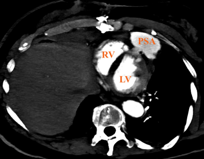
Patients that incur myocardial disruption from penetrating cardiac injuries have an average 6%-10% expectancy rate of reaching the hospital alive. If prompt recognition on arrival is not immediate, the morbidity and mortality are significantly higher due to the secondary physiologic sequalae of either cardiogenic or hemorrhagic shock. Even after a triumphant arrival at a medical facility, out of that 6%-10%, half of those patients are not expected to survive. The unique significance of the presenting case breaks this tradition, expanding past the paradigms and issuing an exceptional understanding of the protective effects that cardiac surgery can futuristically cause through preformed adhesions. In our case, the cardiac adhesions achieved this by containing a penetrating cardiac injury that had caused complete ventricular disruption.

Keywords: Cardiac surgery; Cardiothoracic imaging; Diagnostic radiology; Emergency radiology; Pericardial adhesions; Ventricular pseudoaneurysm.


Figures

Fig 1
Fig. 1
Sagittal Chest CT emphasizing the penetrating trajectory of the knife, coursing posteriorly above the left fifth rib and through the anterior wall of the left ventricle (LV). The penetrating wound traveling from the anterior chest wall to the left ventricle measured 10 cm in length. The large ventricular pseudoaneurysm (PSA) communicates with the left ventricle (LV).
Fig 2
Fig. 2
Axial MIP Chest CT identifying the communicating defect of the contained pseudoaneurysm (PSA) involving the right ventricle (RV) and left ventricle (LV). The violation to the right ventricle measured roughly 0.7 × 0.5 × 0.2 cm and an estimated 0.7 × 1.1 × 0.2 cm defect at the left wall. The body of the pseudoaneurysm measured 6.7 × 3.1 × 3.5 cm.
Fig 3
Fig. 3
Transthoracic apical 4-chamber echocardiogram with color Doppler flow across the anterior right and left ventricular walls, centering over the interventricular septum and emptying into the ventricular pseudoaneurysm (PSA).

References

    1. Restrepo CS, Gutierrez FR, Marmol-Velez JA, Ocazionez D, Martinez-Jimenez S. Imaging patients with cardiac trauma. Radiographics. 2012;32(3):633–649. doi: 10.1148/rg.323115123. Erratum in: Radiographics. 2012 Jul-Aug;32(4):1258. PMID: 22582351. - DOI - PubMed
    1. Warrington SJ, Mahajan K. StatPearls [Internet] StatPearls Publishing; Treasure Island (FL): 2022. Cardiac trauma.
    1. Asfaw I, Arbulu A. Penetrating wounds of the pericardium and heart. Surg Clin North Am. 1977;57(1):37–48. doi: 10.1016/s0039-6109(16)41132-1. - DOI - PubMed
    1. Enders GC, Graeber GM, Poirier RA. Wounds traversing two or more cardiac chambers. Case presentation of two survivors and review of the literature. J Thorac Cardiovasc Surg. 1978;76(1):83–89. - PubMed
    1. Valente T, Bocchini G, Rossi G, Sica G, Davison H, Scaglione M. MDCT prior to median re-sternotomy in cardiovascular surgery: our experiences, infrequent findings and the crucial role of radiological report. Br J Radiol. 2019;92(1101) doi: 10.1259/bjr.20170980. - DOI - PMC - PubMed

Publication types

LinkOut - more resources