. 2023 May 21;15(5):350.
doi: 10.3390/toxins15050350.

Phylogeny-Related Variations in Venomics: A Test in a Subset of Habu Snakes (Protobothrops)

Affiliations

Phylogeny-Related Variations in Venomics: A Test in a Subset of Habu Snakes (Protobothrops)

Hong-Yan Zhao et al. Toxins (Basel). .

Abstract

We conducted a comparative analysis to unveil the divergence among venoms from a subset of Old World habu snakes (Protobothrops) in terms of venomic profiles and toxicological and enzymatic activities. A total of 14 protein families were identified in the venoms from these habu snakes, and 11 of them were shared among these venoms. The venoms of five adult habu snakes were overwhelmingly dominated by SVMP (32.56 ± 13.94%), PLA2 (22.93 ± 9.26%), and SVSP (16.27 ± 4.79%), with a total abundance of over 65%, while the subadult P. mangshanensis had an extremely low abundance of PLA2 (1.23%) but a high abundance of CTL (51.47%), followed by SVMP (22.06%) and SVSP (10.90%). Apparent interspecific variations in lethality and enzymatic activities were also explored in habu snake venoms, but no variations in myotoxicity were found. Except for SVSP, the resemblance of the relatives within Protobothrops in other venom traits was estimated to deviate from Brownian motion evolution based on phylogenetic signals. A comparative analysis further validated that the degree of covariation between phylogeny and venom variation is evolutionarily labile and varies among clades of closely related snakes. Our findings indicate a high level of interspecific variation in the venom proteomes of habu snakes, both in the presence or absence and the relative abundance of venom protein families, and that these venoms might have evolved under a combination of adaptive and neutral mechanisms.

Keywords: Protobothrops; biochemical activity; interspecific variation; phylogenetic signal; venom proteome.


Conflict of interest statement

The authors declare no conflict of interest.

Figures

Figure 1
Figure 1
Major geographical distribution regions of the six habu snakes in the current study. The distribution areas of these snakes were illustrated by ArcGIS based on the data from the GBIF website and complied with the detailed descriptions in “Snakes of China” edited by Ermi Zhao [40]. The animal images were photographed by Jian-Fang Gao; the P. mangshanensis was a subadult specimen and the other habu snakes were adult specimens. A high-resolution image can be found in Figure S1.
Figure 2
Figure 2
The venom proteomic profiles of six habu snakes. The venom proteins of P. cornutus (A), P. jerdonii (B), P. mangshanensis (C), P. mucrosquamatus (D), P. sieversorum (E), and P. xiangchengensis (F) were fractionated on a C18 column as described in the Materials and Methods. The RP-HPLC eluted fractions were collected and separated by SDS-PAGE under reducing conditions (on the left of panels). Protein bands from gels were digested with trypsin and identified by MALDI-TOF/TOF-MS and nESI-MS/MS. The details of the sequenced peptides and assigned protein families are listed in Supplementary Tables S1–S6. The pie charts (on the right of panels) display the relative abundance of the toxin families in the relevant venoms of habu snakes. The venom of P. mangshanensis was collected from subadult specimens, and the venom of the other habu snakes were collected from adult specimens. SVMP, snake venom metalloproteinase; SVSP, snake venom serine proteinase; PLA2, phospholipase A2; CRISP, cysteine-rich secretory protein; BPP/CNP, bradykinin-potentiating and C-type natriuretic peptides; CTL, C-type lectin; LAAO, L-amino acid oxidase; VEGF, vascular endothelial growth factor; NGF, nerve growth factor; PDE, phosphodiesterase; 5′NT, 5′-nucleotidase; PLB, phospholipase B; QC, glutaminyl-peptide cyclotransferase. A high-resolution image can be found in Figure S2.
Figure 2
Figure 2
The venom proteomic profiles of six habu snakes. The venom proteins of P. cornutus (A), P. jerdonii (B), P. mangshanensis (C), P. mucrosquamatus (D), P. sieversorum (E), and P. xiangchengensis (F) were fractionated on a C18 column as described in the Materials and Methods. The RP-HPLC eluted fractions were collected and separated by SDS-PAGE under reducing conditions (on the left of panels). Protein bands from gels were digested with trypsin and identified by MALDI-TOF/TOF-MS and nESI-MS/MS. The details of the sequenced peptides and assigned protein families are listed in Supplementary Tables S1–S6. The pie charts (on the right of panels) display the relative abundance of the toxin families in the relevant venoms of habu snakes. The venom of P. mangshanensis was collected from subadult specimens, and the venom of the other habu snakes were collected from adult specimens. SVMP, snake venom metalloproteinase; SVSP, snake venom serine proteinase; PLA2, phospholipase A2; CRISP, cysteine-rich secretory protein; BPP/CNP, bradykinin-potentiating and C-type natriuretic peptides; CTL, C-type lectin; LAAO, L-amino acid oxidase; VEGF, vascular endothelial growth factor; NGF, nerve growth factor; PDE, phosphodiesterase; 5′NT, 5′-nucleotidase; PLB, phospholipase B; QC, glutaminyl-peptide cyclotransferase. A high-resolution image can be found in Figure S2.
Figure 3
Figure 3
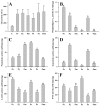
Myotoxicity and enzymatic activities of venoms from habu snakes. C, saline; Pc, P. cornutus; Pj, P. jerdonii; Pm, P. mucrosquamatus; Ps, P. sieversorum; Px, P. xiangchengensis; Pms, P. mangshanensis. Data are expressed as mean value ± SD ((A): n = 4; (BF): n = 3). The enzymatic activities of P. mangshanensis venom (indicated with “*” above the column) were excluded from the statistical analysis due to the venom having been collected from subadult specimens. The significance level is set at α = 0.05, a > b > c > d > e. A high-resolution image can be found in Figure S3.
Figure 4
Figure 4
Local symptoms in the gastrocnemius muscle of the mice after injection of (A) saline; (B) P. cornutus; (C) P. jerdonii; (D) P. mucrosquamatus; (E) P. sieversorum; (F) P. xiangchengensis; (G) P. mangshanensis venoms. High- resolution image can be found in Figure S4.

References

    1. Calvete J.J., Escolano J., Sanz L. Snake venomics of Bitis Species reveals large intragenus venom toxin composition variation: Application to taxonomy of congeneric taxa. J. Proteom. Res. 2007;6:2732–2745. doi: 10.1021/pr0701714. - DOI - PubMed
    1. Casewell N.R., Wüster W., Vonk F.J., Harrison R.A., Fry B.G. Complex cocktails: The evolutionary novelty of venoms. Trends Ecol. Evol. 2013;28:219–229. doi: 10.1016/j.tree.2012.10.020. - DOI - PubMed
    1. Daltry J.C., Wuster W., Thorpe R.S. Diet and snake venom evolution. Nature. 1996;379:537–540. doi: 10.1038/379537a0. - DOI - PubMed
    1. Barlow A., Pook C.E., Harrison R.A., Wüster W. Coevolution of diet and prey-specific venom activity supports the role of selection in snake venom evolution. Proc. R. Soc. B. 2009;276:2443–2449. doi: 10.1098/rspb.2009.0048. - DOI - PMC - PubMed
    1. Healy K., Carbone C., Jackson A.L. Snake venom potency and yield are associated with prey-evolution, predator metabolism and habitat structure. Ecol. Lett. 2019;22:527–537. doi: 10.1111/ele.13216. - DOI - PubMed

Publication types

LinkOut - more resources