Review
. 2023 May 12;10(5):347.
doi: 10.3390/vetsci10050347.

Chronic Progressive Lymphedema in Belgian Draft Horses: Understanding and Managing a Challenging Disease

Affiliations
Review

Chronic Progressive Lymphedema in Belgian Draft Horses: Understanding and Managing a Challenging Disease

Marieke Brys et al. Vet Sci. .

Abstract

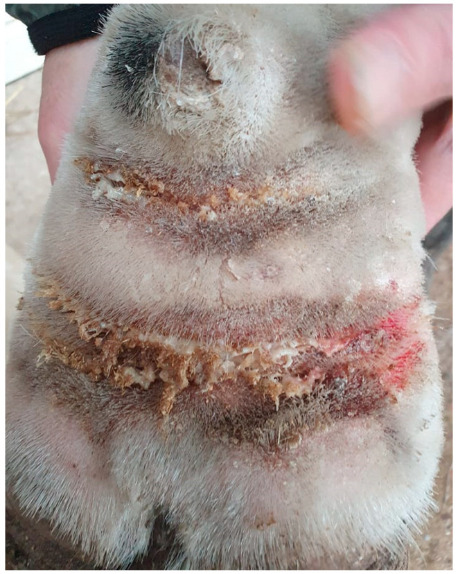
Chronic progressive lymphedema (CPL) in draft horses is characterized by increased dermal thickness and fibrosis, with the development of skinfolds and nodules, hyperkeratosis, and ulcerations on the distal limbs of affected horses. Secondary bacterial, fungal, or parasitic infections frequently complicate and aggravate the lesions, as well as the progression of this disease. CPL has a particularly high prevalence of up to 85.86% in the Belgian draft horse breed. Due to the disease's progressive and incurable nature, affected horses are often euthanized prematurely. The treatment options are solely symptomatic, aimed at improving the horse's quality of life. Despite the severity of this condition, many uncertainties about its etiology and pathogenesis still remain to date. The established scientific research on CPL is rather limited, although there is an urgent need for strategies to tackle this disease. This review summarizes the available knowledge, serving as a guideline for practitioners, and provides perspectives for future research programs.

Keywords: Belgian draft horse; clinical signs; elastin; fibrosis; hyperkeratosis; lymphedema; pathogenesis; skinfolds; treatment.


Conflict of interest statement

The authors declare no conflict of interest.

Figures

Figure 1
Figure 1
Typical lesions of CPL on the distal legs of a Belgian draft horse. Characterizing skinfolds and nodules may be observed on the distal legs, associated with scaling of the skin (after clipping of the feathering).
Figure 2
Figure 2
The vicious cycle of CPL. All factors associated with CPL are interconnected and reinforce each other, with the initiating or causative factor in this cycle still remaining speculative. A genetic factor is thought to play a significant role in the development of CPL, although it could not be identified to date. This genetic predisposition may be present in any of the elements of the vicious cycle.
Figure 3
Figure 3
Dermal and subcutaneous edema of the distal limbs. (a) In the early stages of CPL, localized onset of ‘pitting’ edema can be observed in the fetlock and pastern regions (green arrow). In this horse, slight folding of the skin on the plantar aspect of the cannon bone is also visible. Insert, contralateral leg with absence of edema. (b) Generalized edema of the distal right forelimb. Notice skin fissures on the carpus. (c) Subcutaneous and dermal edema at the level of the palmar region of the distal forelimb in a yearling draft horse.
Figure 4
Figure 4
Skinfolds, the main feature of CPL in the Belgian draft horse. (a) Early-stage lesion of CPL: two folds in the pastern region (green arrow). (b) Advanced stage of CPL: expansion of the folds to the palmar aspect of the cannon bone. (c) Final stage of CPL: skinfolds appear dorsally on the distal limb.
Figure 5
Figure 5
Skinfolds in the neck of a Belgian draft horse. A 6-year-old Belgian draft horse stallion exhibiting folding of the skin in the neck region. The mane has been partially clipped to facilitate easier braiding during studbook inspections (Courtesy of T. van der Weerden, 2021).
Figure 6
Figure 6
Skin fissures on the carpus and tarsus. (a) Typical skin fissure on the palmar aspect of the carpus (green arrow), also referred to as ‘mallenders’. (b) A linear scaling and hyperkeratotic lesion on the lateral aspect of the right hind limb, resulting from a persistent exudative lesion on the dorsal aspect of the tarsus (‘sallenders’).
Figure 7
Figure 7
Verrucous pastern dermatitis. (a) Wart-like nodules (green arrow) in the pastern region of a Belgian draft horse mare. (b) The skin in these areas is highly sensitive to trauma, and consequently poses a high risk of infection and necrosis (Courtesy of F. May, 2021).
Figure 8
Figure 8
Poor hoof horn quality and prominent chestnuts and ergots in draft horses with CPL. (a) The hoof wall exhibits poor keratinization, with visible signs of proliferation, scaling and rings of hoof growth (Courtesy of D. Conder Emge, 2021) (b) Hyperplastic and hyperkeratotic chestnut and (c) ergot in Belgian draft horses.
Figure 9
Figure 9
Cellulitis/lymphangitis in a Belgian draft horse with CPL. (a) Plantar view and (b) medial view of the right hind limb of a CPL-affected draft horse, displaying a ‘stovepipe’ appearance, which can be observed in both lymphangitis and cellulitis (Courtesy of A. Callens, 2022).
Figure 10
Figure 10
Superficial pyoderma in a horse with CPL. The skin displays characteristic signs of bacterial infection, including inflammation, erythema, and exudate (Courtesy of B. Young, 2021).
Figure 11
Figure 11
Intertrigo or skinfold dermatitis in a CPL-affected horse. The skin between the folds is ulcerated, erythematous, and covered with a serous to suppurative exudate (Courtesy of V. Clifton, 2022).
Figure 12
Figure 12
Clinical signs of mange in draft horses. (a) Alopecia is a characteristic feature of mite infestation, commonly observed on the lateral or medial aspect of the cannon bone. (b) Due to intense pruritus, the horse may exacerbate the condition by vigorously scratching or rubbing the affected area, leading to self-induced wounds on the medial side of both the front and hind limbs. (c) An extremely scaly patch of skin as a result of a severe mite infestation. Note the typical white debris surrounding the hair shafts.
Figure 13
Figure 13
Histology of the skin from the pastern region of a CPL-affected Belgian draft horse and a non-affected Belgian draft horse, both aged 15 years (hematoxylin and eosin, magnification ×12.5). The skin of a CPL-affected draft horse (left) is characterized by hyperplasia of the epidermis and hyperkeratosis (yellow asterisk). In the dermis, extensive fibrosis is present (green asterisk). The overall thickness of the skin is significantly increased compared with the skin of a non-affected Belgian draft horse (right).
Figure 14
Figure 14
CPL scoring system, based on the presence and distribution of skinfolds and nodules. N: Normal limb, no clinical signs of CPL. A: Mild to moderate skinfolds and/or nodules in the pastern region, below the fetlock, in which the outline of the pastern maintains a concave shape. A-: Thick skinfolds and/or nodules in the pastern region, below the fetlock, in which the pastern cavity is completely filled with skinfolds and/or nodules. B: Mild to moderate skinfolds and/or nodules in the pastern region, including the fetlock, in which the outline of the pastern maintains a concave shape. B-: Thick skinfolds and/or nodules in the pastern region, including the fetlock, in which the pastern cavity is completely filled with skinfolds and/or nodules. C: Mild to moderate skinfolds and/or nodules in the pastern, fetlock, and metacarpal/metatarsal region, in which the outline of the pastern maintains a concave shape. C-: Thick skinfolds and/or nodules in the pastern, fetlock, and metacarpal/metatarsal region, in which the pastern cavity is completely filled with skinfolds and/or nodules. Xd: Presence of skinfolds and/or nodules on the dorsal side of the limb. This can be present in any of the previously mentioned scores, resulting in Ad(-), Bd(-), or Cd(-).
Figure 15
Figure 15
Radiographic images of the distal legs in CPL-affected horses. (a) A non-affected Belgian draft horse, displaying no skinfolds. (Courtesy of M. Oosterlinck and K. Vanderperren, 2023). (b) A moderately affected Gypsy Cob mare with clear skinfolds in the pastern region and some slight folding of the skin plantar to the cannon bone. (Courtesy of E. Evrard, 2021) (c) A severely affected draft horse with pronounced, thick skinfolds on the overall distal limb. (Courtesy of Boschhoven paard, 2019).
Figure 16
Figure 16
Diathermy surgery in a Gypsy Cob with CPL and warty-like nodules. (a) Right distal forelimb before surgery. (b) Right distal forelimb 12 days after surgery. (c) Regrowth appeared within seven months after surgery. (Courtesy of A. Torstensson, 2022).
Figure 17
Figure 17
Effects of hemorrhoid cream treatment in a Gypsy Cob with mild CPL and warty-like nodules. (a) Palmar view of the forelimbs prior to treatment. Note the presence of a high number of wart-like growths in the pastern region and fetlock. (b) Palmar view of the forelimbs after treatment for six months with hemorrhoid cream. The growths have resolved almost completely. (Courtesy of H. Lane and H. Johnsen, 2022).

References

    1. Schutzer E. Ph.D. Thesis. Medizinische Fakultät der Universität Leipzig; Leipzig, Germany: 1909. Ein Beitrag zur Kenntnis der Dermatitis Chronica Verrucosa (Sogenannter Straubfuss)
    1. Gustine G. Ph.D. Thesis. Vereinigte Medizinische Fakultät der Groβherzöglichen Hessischen Ludwigs-Universität zu Gieβen; Gieβen, Germany: 1910. Die Sogenannte Warzenmauke des Pferdes “Dermatitis Chronica Verrucosa”.
    1. Wussow W., Hartwig W. Die Zuchtverwendungsdauer der Beschäler des Landgestüt Kreuz und ihre Abgangsursachen. Tierzucht. 1954;10:336–342.
    1. Ferraro G.L. Chronic progressive lymphedema in draft horses. J. Equine Vet. Sci. 2003;23:189–190. doi: 10.1053/jevs.2003.56. - DOI
    1. Affolter V.K. Chronic Progressive Lymphedema in Draft Horses. Vet. Clin. N. Am. Equine Pract. 2013;29:589–605. doi: 10.1016/j.cveq.2013.08.007. - DOI - PubMed

LinkOut - more resources