. 2023 Jun 13;95(23):8747-8751.
doi: 10.1021/acs.analchem.3c00958. Epub 2023 May 26.

Shaping Nanobodies and Intrabodies against Proteoforms

Affiliations

Shaping Nanobodies and Intrabodies against Proteoforms

Bojana Leonard et al. Anal Chem. .

Abstract

Proteoforms expand genomic diversity and direct developmental processes. While high-resolution mass spectrometry has accelerated characterization of proteoforms, molecular techniques working to bind and disrupt the function of specific proteoforms have lagged behind. In this study, we worked to develop intrabodies capable of binding specific proteoforms. We employed a synthetic camelid nanobody library expressed in yeast to identify nanobody binders of different SARS-CoV-2 receptor binding domain (RBD) proteoforms. Importantly, employment of the positive and negative selection mechanisms inherent to the synthetic system allowed for amplification of nanobody-expressing yeast that bind to the original (Wuhan strain RBD) but not the E484 K (Beta variant) mutation. Nanobodies raised against specific RBD proteoforms were validated by yeast-2-hybrid analysis and sequence comparisons. These results provide a framework for development of nanobodies and intrabodies that target proteoforms.


Conflict of interest statement

The authors declare no competing financial interest.

Figures
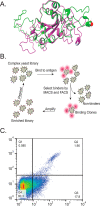
Figure 1
Figure 1
(A) AlphaFold predicted structure of the Wuhan SARS-CoV-2 Spike RBD (in green) with the E residue labeled as spheres on the green protein and the 484 K RBD (in pink) with the K residue labeled as a sphere. Notice the predicted changes in the Beta sheet and alpha helical content as well as the position of the disordered loop region between the two proteoforms. (B) Schematic representation of negative and positive selections of different RBD proteoforms through multiple rounds of MACS and FACS. (C) Final FACS analyses of yeast that were positively selected for E484 K RBD proteoforms (incubated here with E484 K RBD-A647 and Wuhan RBD-A488). The FACS shows a clear population of E484-only binding yeast (Q3) and yeast that bind to both the Wuhan and E484 K RBD proteoforms (Q2).
Figure 2
Figure 2
Analysis of nanobody populations from yeast surface display enrichment. (A) Venn diagram analysis of Nb populations trained to bind to either the Wuhan or the E484 K RBD proteoform. (B) Heatmap of yeast nanobodies trained to bind either the Wuhan, E484 K, or both RBD proteoforms. Red areas indicate the presence of a given Nb. Gray areas indicate the absence of a Nb.
Figure 3
Figure 3
(A) Description of yeast-two-hybrid assay for validation of RBD proteoform-binding nanobodies. The RBD antigen (Ag) is fused to the GAL-4 transcription factor DNA binding domain (BD) to make an Ag-BD fusion protein. The pool of surface-display-enriched Nbs were cloned in-frame with the GAL-4 transcription factor activation domain (AD) to make a Nb-AD fusion. Haploid yeast carrying both fusion protein types were mated to produce a diploid yeast test for growth selection on SC-HIS media (with and without 3-AT). Yeast were also grown in the absence of SC-Leu and SC-Trp to select for the presence of the GAL-4 fusion plasmids. (B) (Left) Y2H growth analysis on SC-Trp and SC-Leu media (selects for the presence of the GAL-4 fusion plasmids). (Right) Y2H growth analysis on SC-Trp, SC-Leu, and SC-HIS media (selects for interactions between the GAL4-BD and GAL4-AD fusions). Labeling key: Wuhan (Wu) Nb + Blank = negative control with the Wuhan RBD-trained Nb library mated to the GAL-4 DNA binding domain (absent an RBD proteoform fusion). Wuhan Nb + Wuhan RBD = Nbs trained against the Wuhan RBD mated to the Wuhan RBD in the Y2H. Wuhan Nb + E484 K RBD = Nbs trained against the Wuhan RBD mated against the E484 K RBD. Wuhan Nb + E484Q RBD = Nbs trained against the Wuhan RBD mated against the E484Q RBD. E484 K Nb + Blank = negative control with the E484 K RBD-trained Nb library mated to the GAL-4 DNA binding domain (absent an RBD proteoform). E484 K Nb + Wuhan RBD = Nbs trained against the E484 K RBD mated to the Wuhan RBD. E484k Nb + E484 K RBD = Nbs trained against the E484 K RBD mated to the E484 K RBD. E484 K Nb + E484Q RBD = Nbs trained against the E484 K RBD mated to the E484Q RBD. The dilution series of the yeast is indicated below each growth plate.

References

    1. Smith L. M; Kelleher N. L Proteoform: a single term describing protein complexity. Nat. Methods 2013, 10 (3), 186–187. 10.1038/nmeth.2369. - DOI - PMC - PubMed
    1. Adams L. M.; DeHart C. J.; Drown B. S.; Anderson L. C.; Bocik W.; Boja E. S.; Hiltke T. M.; Hendrickson C. L.; Rodriguez H.; Caldwell M.; Vafabakhsh R.; Kelleher N. L. Mapping the KRAS proteoform landscape in colorectal cancer identifies truncated KRAS4B that decreases MAPK signaling. J. Biol. Chem. 2023, 299 (1), 102768.10.1016/j.jbc.2022.102768. - DOI - PMC - PubMed
    1. Ntai I.; Fornelli L.; DeHart C. J.; Hutton J. E.; Doubleday P. F.; LeDuc R. D.; van Nispen A. J.; Fellers R. T.; Whiteley G.; Boja E. S.; Rodriguez H.; Kelleher N. L. Precise characterization of KRAS4b proteoforms in human colorectal cells and tumors reveals mutation/modification cross-talk. Proc. Natl. Acad. Sci. U. S. A. 2018, 115 (16), 4140–4145. 10.1073/pnas.1716122115. - DOI - PMC - PubMed
    1. Melani R. D.; Gerbasi V. R.; Anderson L. C.; Sikora J. W.; Toby T. K.; Hutton J. E.; Butcher D. S.; Negrao F.; Seckler H. S.; Srzentic K.; Fornelli L.; Camarillo J. M.; LeDuc R. D.; Cesnik A. J.; Lundberg E.; Greer J. B.; Fellers R. T.; Robey M. T.; DeHart C. J.; Forte E.; Hendrickson C. L.; Abbatiello S. E.; Thomas P. M.; Kokaji A. I.; Levitsky J.; Kelleher N. L. The Blood Proteoform Atlas: A reference map of proteoforms in human hematopoietic cells. Science 2022, 375 (6579), 411–418. 10.1126/science.aaz5284. - DOI - PMC - PubMed
    1. Toby T. K.; Abecassis M.; Kim K.; Thomas P. M.; Fellers R. T.; LeDuc R. D.; Kelleher N. L.; Demetris J.; Levitsky J. Proteoforms in Peripheral Blood Mononuclear Cells as Novel Rejection Biomarkers in Liver Transplant Recipients. Am. J. Transplant 2017, 17 (9), 2458–2467. 10.1111/ajt.14359. - DOI - PMC - PubMed

Publication types

Substances

Supplementary concepts