. 2023 May 26;9(21):eadf0133.
doi: 10.1126/sciadv.adf0133. Epub 2023 May 26.

LOXL4, but not LOXL2, is the critical determinant of pathological collagen cross-linking and fibrosis in the lung

Affiliations

LOXL4, but not LOXL2, is the critical determinant of pathological collagen cross-linking and fibrosis in the lung

Hsiao-Yen Ma et al. Sci Adv. .

Abstract

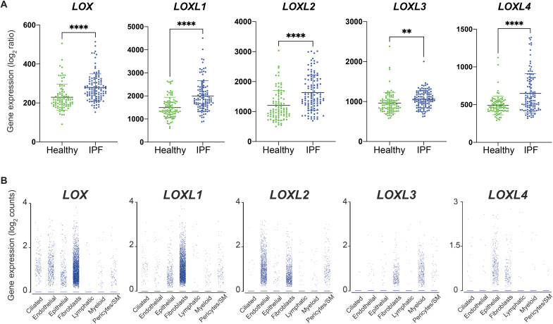
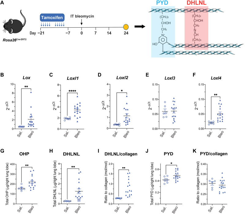
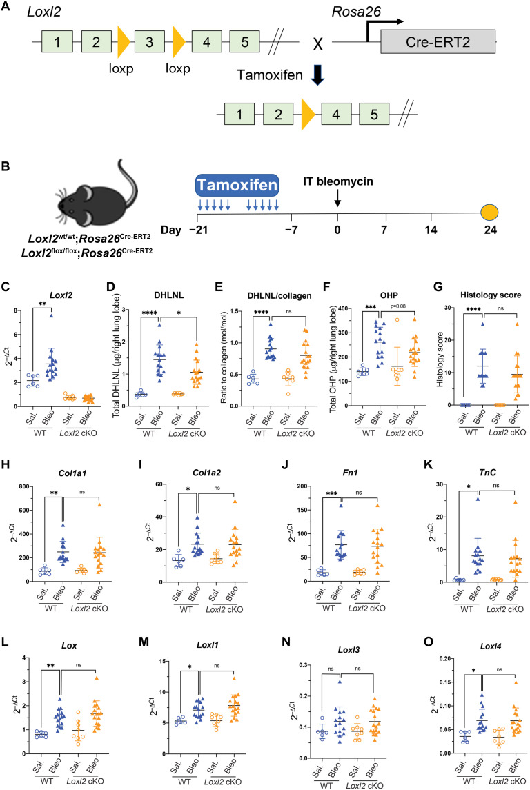
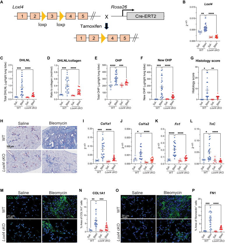
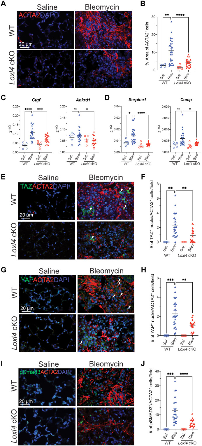
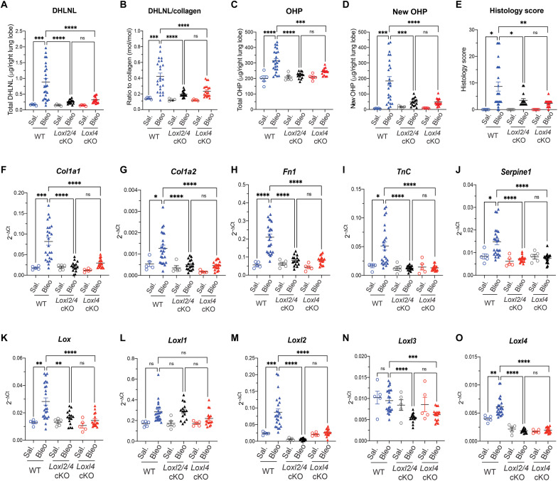
Idiopathic pulmonary fibrosis is a progressive fibrotic disease characterized by excessive deposition of (myo)fibroblast produced collagen fibrils in alveolar areas of the lung. Lysyl oxidases (LOXs) have been proposed to be the central enzymes that catalyze the cross-linking of collagen fibers. Here, we report that, while its expression is increased in fibrotic lungs, genetic ablation of LOXL2 only leads to a modest reduction of pathological collagen cross-linking but not fibrosis in the lung. On the other hand, loss of another LOX family member, LOXL4, markedly disrupts pathological collagen cross-linking and fibrosis in the lung. Furthermore, knockout of both Loxl2 and Loxl4 does not offer any additive antifibrotic effects when compared to Loxl4 deletion only, as LOXL4 deficiency decreases the expression of other LOX family members including Loxl2. On the basis of these results, we propose that LOXL4 is the main LOX activity underlying pathological collagen cross-linking and lung fibrosis.


Figures

Fig. 1.
Fig. 1.. Expression of LOX family members in human IPF lungs.
(A) Expression of LOX, LOXL1, LOXL2, LOXL3, and LOXL4 in healthy and IPF human lungs. (B) Expression of LOX, LOXL1, LOXL2, LOXL3, and LOXL4 in individual cell type in human IPF lungs. SM, smooth muscle cells. Data represent means ± SD. **P < 0.01 and ****P < 0.0001. P value is calculated using one-way ANOVA.
Fig. 2.
Fig. 2.. Collagen cross-linking and Lox/Loxl expression in the bleomycin model.
(A) Schematic regime of Rosa26Cre-ERT2 mice under intratracheal (IT) bleomycin challenge. Lungs were harvested for terminal analyses on day 24 after bleomycin challenge. (B to F) mRNA expression of Lox family members from lung tissue. (G) Lung total OHP. (H to K) Quantification of lung collagen cross-linking by (H) total DHLNL, (I) DHLNL normalized by collagen, (J) total PYD, and (K) PYD normalized by collagen. Saline, n = 6; bleomycin, n = 16. Data represent means ± SD. *P < 0.05, **P < 0.01, and ****P < 0.0001. P value is calculated using unpaired, two-tailed t test (B) to (K).
Fig. 3.
Fig. 3.. LOXL2 modestly contributes to pathological collagen cross-linking but not fibrosis in the bleomycin model.
(A) Schematic conditional Loxl2 targeting. The exon 3 of Loxl2 gene has been flanked by loxP site. The Loxl2flox/flox mice breed with Rosa26Cre-ERT2 mice to generate Loxl2 cKO mice (Loxl2flox/flox; Rosa26Cre-ERT2) where Cre promoted recombination can be induced by tamoxifen. (B) Schematic regime of tamoxifen-induced Loxl2 deletion followed by intratracheal bleomycin challenge. Lungs were harvested for terminal analyses on day 24 after bleomycin challenge. (C) mRNA expression shows the efficient deletion of Loxl2 in the lungs from cKO mice. (D and E) Quantification of lung collagen cross-linking by (D) total DHLNL and (E) DHLNL normalized by collagen. (F and G) Quantification of lung fibrosis by (F) total OHP and (G) histology score. (H to K) mRNA expression of selective profibrotic genes from lung tissue. (L to O) mRNA expression of Lox family members from lung tissue. WT: saline, n = 6; bleomycin, n = 15; Loxl2 cKO: saline, n = 8; bleomycin, n = 17. Data represent means ± SD. *P < 0.05, **P < 0.01, and ****P < 0.0001. P value is calculated using one-way analysis of variance (ANOVA). ns, not significant.
Fig. 4.
Fig. 4.. Loxl4 cKO mice are protective against bleomycin-induced lung fibrosis.
(A) Schematic conditional Loxl4 targeting. The exon 3 of Loxl4 gene has been flanked by loxP site. The Loxl4flox/flox mice breed with Rosa26Cre-ERT2 mice to generate Loxl4 cKO mice (Loxl4flox/flox; Rosa26Cre-ERT2) where Cre promoted recombination can be induced by tamoxifen. (B) mRNA expression shows the efficient deletion of Loxl4 in the lungs from cKO mice. (C and D) Quantification of lung collagen cross-linking by (C) total DHLNL and (D) DHLNL normalized by collagen. (E and F) Quantification of lung fibrosis by (E) total OHP and (F) newly synthesized OHP. (G) Histology score of lung fibrosis lesion. (H) Representative images of trichrome staining of lung fibrosis. (I to L) mRNA expression of selective profibrotic genes from lung tissue. (M and N) (M) Representative immunofluorescent images of COL1A1 in lung tissue with quantification of (N) percentage of COL1A1+ area. (O and P) (O) Representative immunofluorescent images of FN1 in lung tissue with quantification of (P) percentage of FN1+ area. WT: saline, n = 5; bleomycin, n = 24; Loxl4 cKO: saline, n = 5; bleomycin, n = 17. Data represent means ± SD. *P < 0.05, **P < 0.01, ***P < 0.001, and ****P < 0.0001. P value is calculated using one-way ANOVA. DAPI, 4′,6-diamidino-2-phenylindole.
Fig. 5.
Fig. 5.. LOXL4 deficiency disrupts TGFβ signaling and inactivates myofibroblasts in the bleomycin injured lungs.
(A and B) (A) Representative immunofluorescent images of ACTA2 in lung tissue with quantification of (B) percentage of ACTA2+ area. (C and D) mRNA expression of selective (C) YAP/TAZ target genes and (D) TGFβ target genes from lung tissue. (E and F) (E) Representative immunofluorescent images of TAZ and ACTA2 in lung tissue with quantification of (F) number of TAZ+ nuclei/ACTA2+ cells per field. (G and H) (G) Representative immunofluorescent images of YAP and ACTA2 in lung tissue with quantification of (H) number of YAP+ nuclei/ACTA2+ cells per field. Arrowheads present (E) TAZ+ nuclei/ACTA2+ cells and (G) YAP+ nuclei/ACTA2+ cells. (I and J) (I) Representative immunofluorescent images of pSMAD3 and ACTA2 in lung tissue with quantification of (J) number of pSMAD3+/ACTA2+ cells per field. Data in (B), (F), (H), and (J) represent mean value of five random fields per image. Data represent means ± SD. *P < 0.05, **P < 0.01, ***P < 0.001, and ****P < 0.0001. P value is calculated using one-way ANOVA.
Fig. 6.
Fig. 6.. LOXL2 deficiency has no extra protective effects against bleomycin-induced pathological collagen cross-linking and lung fibrosis in the absence of LOXL4.
(A and B) Quantification of lung collagen cross-linking by (A) total DHLNL and (B) DHLNL normalized by collagen. (C to E) Quantification of lung fibrosis by (C) total OHP, (D) newly synthesized OHP, and (E) histology score of fibrosis lesion. (F to J) mRNA expression of selective profibrotic genes from lung tissue. (K to O) mRNA expression of Lox family members from lung tissue. WT: saline, n = 5; bleomycin, n = 24; Loxl2/4 cKO: saline, n = 5; bleomycin, n = 15; Loxl4 cKO: saline, n = 5; bleomycin, n = 17. Data represent means ± SD. *P < 0.05, **P < 0.01, ***P < 0.001, and ****P < 0.0001. P value is calculated using one-way ANOVA.

References

    1. D. S. Kim, H. R. Collard, T. E. King Jr., Classification and natural history of the idiopathic interstitial pneumonias. Proc. Am. Thorac. Soc. 3, 285–292 (2006). - PMC - PubMed
    1. A. Nalbandian, K. Sehgal, A. Gupta, M. V. Madhavan, C. McGroder, J. S. Stevens, J. R. Cook, A. S. Nordvig, D. Shalev, T. S. Sehrawat, N. Ahluwalia, B. Bikdeli, D. Dietz, C. der-Nigoghossian, N. Liyanage-Don, G. F. Rosner, E. J. Bernstein, S. Mohan, A. A. Beckley, D. S. Seres, T. K. Choueiri, N. Uriel, J. C. Ausiello, D. Accili, D. E. Freedberg, M. Baldwin, A. Schwartz, D. Brodie, C. K. Garcia, M. S. V. Elkind, J. M. Connors, J. P. Bilezikian, D. W. Landry, E. Y. Wan, Post-acute COVID-19 syndrome. Nat. Med. 27, 601–615 (2021). - PMC - PubMed
    1. P. M. George, A. U. Wells, R. G. Jenkins, Pulmonary fibrosis and COVID-19: The potential role for antifibrotic therapy. Lancet Respir. Med. 8, 807–815 (2020). - PMC - PubMed
    1. L. Richeldi, H. R. Collard, M. G. Jones, Idiopathic pulmonary fibrosis. Lancet 389, 1941–1952 (2017). - PubMed
    1. G. Raghu, B. Rochwerg, Y. Zhang, C. A. Garcia, A. Azuma, J. Behr, J. L. Brozek, H. R. Collard, W. Cunningham, S. Homma, T. Johkoh, F. J. Martinez, J. Myers, S. L. Protzko, L. Richeldi, D. Rind, M. Selman, A. Theodore, A. U. Wells, H. Hoogsteden, H. J. Schünemann; American Thoracic Society; European Respiratory society; Japanese Respiratory Society; Latin American Thoracic Association , An official ATS/ERS/JRS/ALAT clinical practice guideline: Treatment of idiopathic pulmonary fibrosis. An update of the 2011 clinical practice guideline. Am. J. Respir. Crit. Care Med. 192, e3–e19 (2015). - PubMed