Review
. 2023 May 25;186(11):2288-2312.
doi: 10.1016/j.cell.2023.04.025.

A 360° view of the inflammasome: Mechanisms of activation, cell death, and diseases

Affiliations
Review

A 360° view of the inflammasome: Mechanisms of activation, cell death, and diseases

Katherine C Barnett et al. Cell. .

Abstract

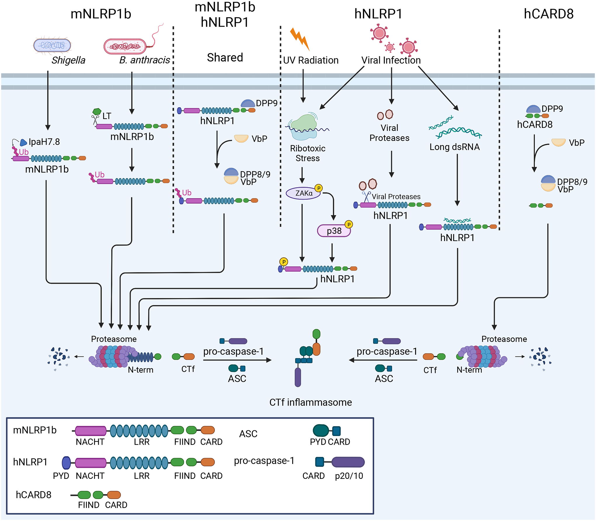
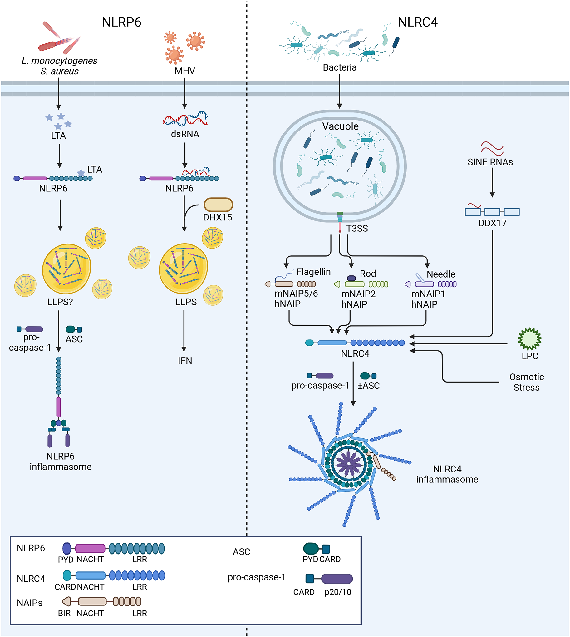
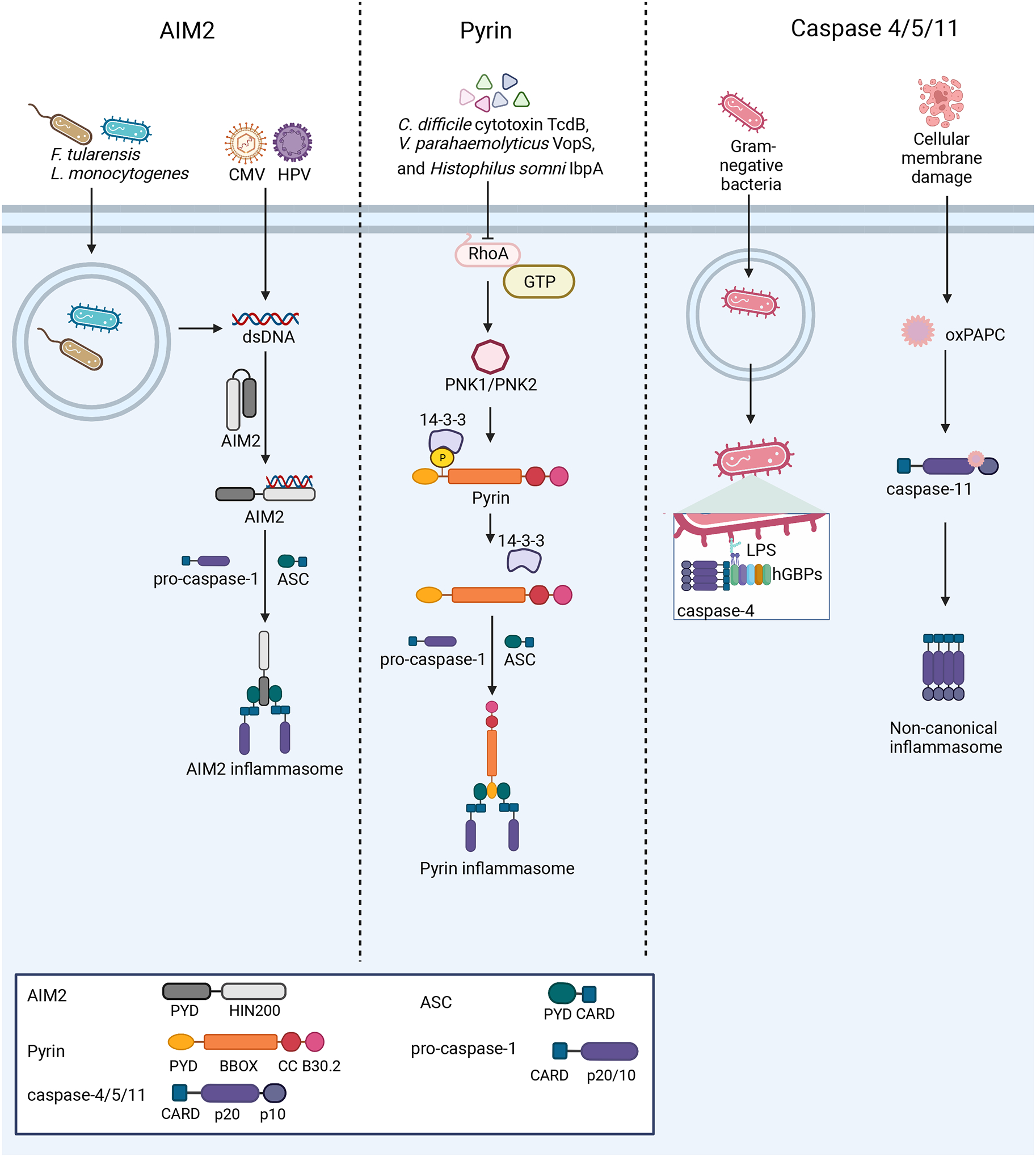
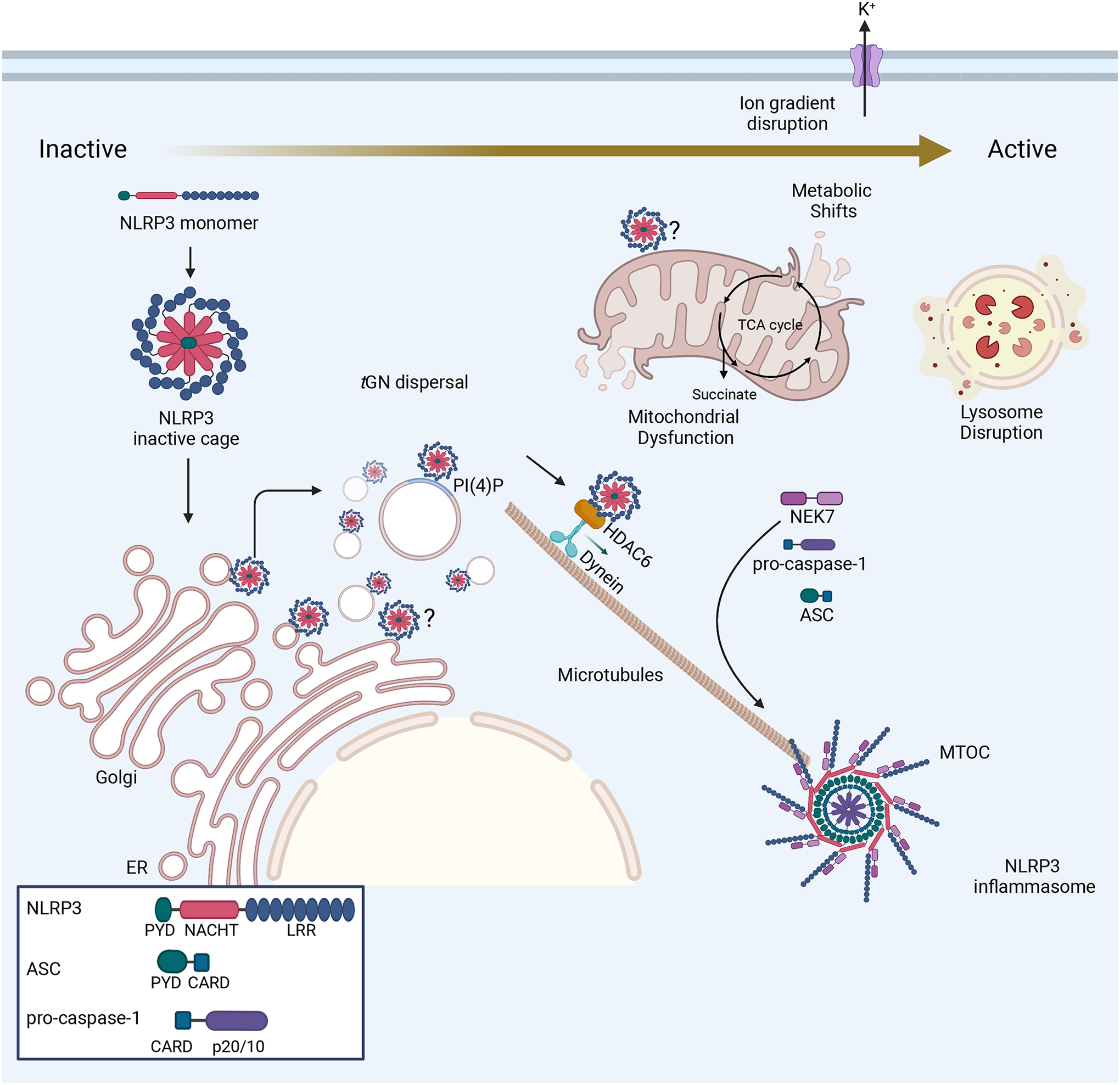
Inflammasomes are critical sentinels of the innate immune system that respond to threats to the host through recognition of distinct molecules, known as pathogen- or damage-associated molecular patterns (PAMPs/DAMPs), or disruptions of cellular homeostasis, referred to as homeostasis-altering molecular processes (HAMPs) or effector-triggered immunity (ETI). Several distinct proteins nucleate inflammasomes, including NLRP1, CARD8, NLRP3, NLRP6, NLRC4/NAIP, AIM2, pyrin, and caspases-4/-5/-11. This diverse array of sensors strengthens the inflammasome response through redundancy and plasticity. Here, we present an overview of these pathways, outlining the mechanisms of inflammasome formation, subcellular regulation, and pyroptosis, and discuss the wide-reaching effects of inflammasomes in human disease.

Keywords: AIM2; CARD8; GSDMD; IL-18; IL-1β; NLRC4; NLRP1; NLRP3; NLRP6; caspase-1; caspase-11; caspase-4; caspase-5; inflammasome; pyrin; pyroptosis.


Conflict of interest statement

Declaration of interests J.P.-Y.T. is a cofounder of IMMvention Therapeutix that works on inflammasome inhibitors.

Figures

Figure 1:
Figure 1:. Distinct and shared mechanisms of murine NLRP1b (mNLRP1b) and human NLRP1 (hNLRP1) and human CARD8.
NLRP1 activation mechanisms vary between specific alleles in the murine population and between species. Some mNLRP1b alleles are sensitive to B. anthracis LT and S. flexneri effector IpaH7.8, which cleave or ubiquitinate mNLRP1b, respectively, instigating proteasomal degradation of the mNLRP1b N-terminus and formation of the mNLRP1b CTf inflammasome. Both mNLRP1b and hNLRP1 respond to DPP8/9 inhibition through Val-boroPro (VbP), leading to Ctf inflammasome formation. hNLRP1, but not mNLRP1b, responds to viral proteases, ribotoxic stress, and viral RNA to promote CTf inflammasome formation. hCARD8 responds to Val-boroPro similarly to mNLRP1b and hNLRP1 and forms CTf inflammasome.
Figure 2:
Figure 2:. Mechanisms of NLRP6 and NLRC4 inflammasome activation
Left: NLRP6 responds to both LTA from bacterial infections and viral RNA from mouse hepatitis virus (MHV). In response to LTA, NLRP6 nucleates an inflammasome, while the response to RNA is mediated by DHX15 and promotes IFN production. LLPS is reported to govern both of these responses. Right: NLRC4 and hNAIP or mNAIP1–6 form inflammasomes upon recognition of flagellin or T3SS components from bacterial infections, as described in the text. More recent reports suggest that NLRC4 may also recognize sterile stimuli, including SINE RNAs through DDX17 activity, osmotic stress, and LPC. NLRC4 inflammasomes may form with or without ASC.
Figure 3:
Figure 3:. Nucleation of the AIM2, pyrin, and non-canonical inflammasomes
Left: AIM2 detects dsDNA from a variety of sources, including bacterial and viral infections. Upon dsDNA binding, AIM2 oligomerizes along the dsDNA and interacts with ASC to form the inflammasome. Center: At steady-state, pyrin is kept in an inactive conformation through phosphorylation by PKN1/2 and interaction of 14-3-3 proteins with this PTM. Several bacterial toxins activate the pyrin inflammasome through inactivation of the RhoA GTPase, which in turn causes pyrin to be dephosphorylated and nucleate inflammasome formation. Right: Murine caspase-11 and human caspases-4/5 act as PRRs. Guanylate-binding proteins (GBPs) assemble on the surface of Gram-negative bacteria after escape from the vacuole, exposing Lipid A moieties from LPS. This GBP coat then serves as a platform for caspase-4 recruitment and activation. The binding of LPS or oxPAPC by these caspases promotes their activation and downstream inflammasome formation.
Figure 4:
Figure 4:. Cellular orchestration of the NLRP3 inflammasome
Prior to activation, NLRP3 exists in an inactivate cage conformation and is reported to associate with many different organelles, including the mitochondria and the ER. Recent work revealed that NLRP3 associates with the TGN through electrostatic interactions with the membrane component lipid PI(4)P,, and a new study describes how these PI(4)P+ vesicles are endosomal in origin, displaying markers of early endosomes and the TGN,. Upon its activation through ion gradient disruption, organelle dysfunction, or metabolic shifts, NLRP3 mediates the dispersal of the TGN and traffics along microtubules in a manner dependent on HDAC6 and dynein. At the MTOC, NLRP3 associates with NEK7, and NLRP3 monomers assemble into a decameric inflammasome.
Figure 5:
Figure 5:. Regulation of cell membrane rupture following inflammasome activation
Left: Caspase-1 cleaves IL-1β/IL-18 and GSDMD to free its N-terminal (GSDMD-NT) fragment. The GSDMD-NT forms a pore on the plasma membrane, releasing IL-1β/IL-18 and disrupting cellular ion gradients to execute pyroptosis. Mitochondrial ROS downstream of Ragulator-Rag-mTORC1 activity regulates GSDMD-NT pore formation and loss of these signals prevents GSDMD NT oligomerization. GSDMD-NT pores may also form on the mitochondria through interaction with cardiolipin, causing the release of mtDNA that can activate cGAS/STING signaling and ROS that can trigger necroptosis. Inflammasomes also promote NINJ1 oligomerization in the plasma to perpetuate membrane rupture and cell death. Right: GSDMD-NT pores can be repaired through ESCRT-III-mediated cell membrane budding, triggered by Ca2+ influx. GSDMD-NT pores may also be repaired through caspase-7 activity. Caspase-7 and ASM are released into the extracellular space, where caspase-7 cleaves ASM to activate it. This causes the enrichment of ceramide on the plasma membrane through ASM activity, which promotes internalization of the microdomains on the plasma membrane that contain GSDMD pores. Both of these mechanisms lead to decreased prevalence of GSDMD pores on the plasma membrane and may explain how some cells do not die following inflammasome activation.

References

    1. Poyet J-L, Srinivasula SM, Tnani M, Razmara M, Fernandes-Alnemri T, and Alnemri ES (2001). Identification of Ipaf, a Human Caspase-1-activating Protein Related to Apaf-1*. J Biol Chem 276, 28309–28313. 10.1074/jbc.c100250200. - DOI - PubMed
    1. Wang L, Manji GA, Grenier JM, Al-Garawi A, Merriam S, Lora JM, Geddes BJ, Briskin M, DiStefano PS, and Bertin J (2002). PYPAF7, a Novel PYRIN-containing Apaf1-like Protein That Regulates Activation of NF-κB and Caspase-1-dependent Cytokine Processing*. J Biol Chem 277, 29874–29880. 10.1074/jbc.m203915200. - DOI - PubMed
    1. Kagan JC, Magupalli VG, and Wu H (2014). SMOCs: supramolecular organizing centres that control innate immunity. Nature Reviews Immunology 14, 821–826. 10.1038/nri3757. - DOI - PMC - PubMed
    1. Martinon F, Burns K, and Tschopp J (2002). The Inflammasome A Molecular Platform Triggering Activation of Inflammatory Caspases and Processing of proIL-β. Mol Cell 10, 417–426. 10.1016/s1097-2765(02)00599-3. - DOI - PubMed
    1. Koonin EV, and Aravind L (2000). The NACHT family - a new group of predicted NTPases implicated in apoptosis and MHC transcription activation. Trends Biochem. Sci 25, 223–224. - PubMed

Publication types

MeSH terms

LinkOut - more resources