. 2023 Jun 14;31(6):928-936.e4.
doi: 10.1016/j.chom.2023.04.015. Epub 2023 Apr 25.

Memory profiles distinguish cross-reactive and virus-specific T cell immunity to mpox

Affiliations

Memory profiles distinguish cross-reactive and virus-specific T cell immunity to mpox

Sarah Adamo et al. Cell Host Microbe. .

Abstract

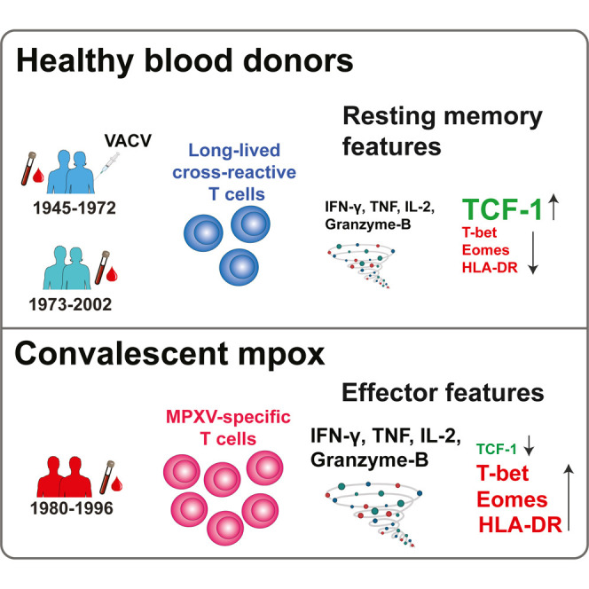
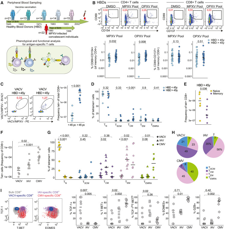
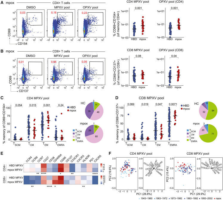
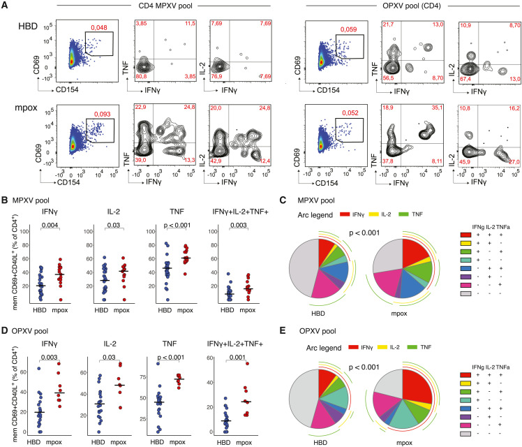
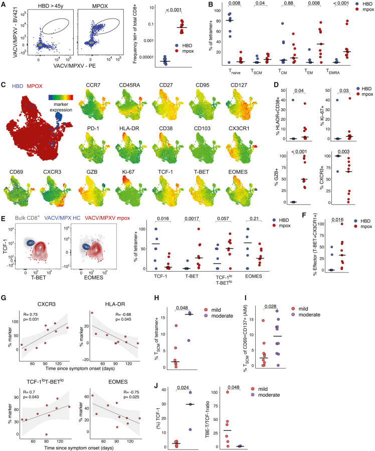
Mpox represents a persistent health concern with varying disease severity. Reinfections with mpox virus (MPXV) are rare, possibly indicating effective memory responses to MPXV or related poxviruses, notably vaccinia virus (VACV) from smallpox vaccination. We assessed cross-reactive and virus-specific CD4+ and CD8+ T cells in healthy individuals and mpox convalescent donors. Cross-reactive T cells were most frequently observed in healthy donors over 45 years. Notably, long-lived memory CD8+ T cells targeting conserved VACV/MPXV epitopes were identified in older individuals more than four decades after VACV exposure and exhibited stem-like characteristics, defined by T cell factor-1 (TCF-1) expression. In mpox convalescent donors, MPXV-reactive CD4+ and CD8+ T cells were more prevalent than in controls, demonstrating enhanced functionality and skewing toward effector phenotypes, which correlated with milder disease. Collectively, we report robust effector memory MPXV-specific T cell responses in mild mpox and long-lived TCF-1+ VACV/MPXV-specific CD8+ T cells decades after smallpox vaccination.

Keywords: MPXV; T cells; VACV; infectious diseases; long-lived memory T cells; monkeypox; mpox; vaccinia virus; viral immunity.


Conflict of interest statement

Declaration of interests M.B. is a consultant for Oxford Immunotec, Mabtech, Bristol-Myers Squibb, and MSD.

Figures

None
Graphical abstract
Figure 1
Figure 1
VACV-induced T cell responses associate with cross-reactive responses to MPXV in HBDs. (A) Schematic of study design. (B) Representative plots showing CD69/CD154 and CD69/CD137 expression after peptide stimulation on memory CD4+ and CD8+ T cells (top) and quantification of net frequencies (background-subtracted using dimethyl sulfoxide [DMSO] control) (bottom). (C) Tetramer staining of VACV/MPXV epitopes in HBDs of different age groups and quantification. (D) Comparison of memory population frequency among tetramer+ CD8+ T cells in different age subgroups. (E) Frequencies of naive and memory T cells within tetramer+ CD8+ T cells from HBDs. (F) Frequencies of VACV+, IAV+, and CMV+ CD8+ T cells in HBDs. (G) Comparison of naive and memory population frequency among tetramer+ CD8+ T cells. (H) Subpopulation distribution of memory VACV+, IAV+, and CMV+ CD8+ T cells in HBDs. (I) Representative plots showing transcription factors expression (left) and frequency of transcription factor and Granzyme B expression within VACV+, IAV+, and CMV+ CD8+ T cells in HBDs (right). In (B)–(G) and (I), Mann-Whitney test. See also Figure S1.
Figure 2
Figure 2
Magnitude of T cell responses in HBDs and mpox convalescent patients (A) Representative flow plots showing CD69/CD154 expression after peptide stimulations, gated on memory CD4+ cells (left) and comparison of net frequencies of AIM+ CD4+ between mpox patients and HBDs (right). (B) Representative flow plots showing CD69/CD137 expression on memory CD8+ T cells and quantification as in (A). (C) Comparison of frequencies (left) and distribution (right) of CD4+ T cell memory subsets in MPXV-reactive cells of HBDs and mpox patients. (D) Comparison of frequencies (left) and distribution (right) of CD8+ T cell memory subsets as in (C). (E) Heatmap showing marker expression level on MPXV-reactive CD4+ and CD8+ T cells between HBDs and convalescent donors. (F) PCA plot using the dataset in (E) to show the segregation of HBDs and convalescent donors and key markers associated with the segregation. In (A)–(E), Mann-Whitney test. p < 0.05, ∗∗p < 0.01, ∗∗∗p < 0.001, and ∗∗∗∗p < 0.0001 in (E). See also Figures S2 and S3.
Figure 3
Figure 3
Functional profile of AIM+ CD4+ T cells (A) Representative flow plot for IFN-γ, IL-2, and TNF expression within the AIM+ CD4+ T cell population following stimulation with MPXV and OPXV peptide pools. (B and C) Comparison between patterns of single expression and co-expression for IFN-γ, IL-2, and TNF after MPXV pool stimulation. (D and E) Comparison between patterns of single expression and co-expression for IFN-γ, IL-2, and TNF after OPXV pool stimulation. In (B) and (D), Mann-Whitney test. In (C) and (E), permutation test.
Figure 4
Figure 4
Phenotypical profile of VACV/MPXV tetramer+ CD8+ T cells in HBDs and mpox patients (A) Representative flow plots showing VACV/MPXV tetramer staining on CD8+ T cells (left) and frequencies (right). (B) Frequencies of naive and memory T cell subsets within tetramer+ CD8+ T cells in HBDs and convalescent donors. (C) UMAP visualization of tetramer+ CD8+ T cells from HBDs and convalescent donors, and expression of individual markers colored by marker expression levels. (D) Frequency of HLA-DR+CD38+, Ki-67+, Granzyme B+ CD8+ T cells, and CXCR3+ expression in HBDs vs. convalescent donors. (E) Representative flow plots of transcription factors expression within VACV/MPXV+ CD8+ T cells in HBDs vs. convalescent donors (left) and quantification (right). (F) Frequency of T-bet+CX3CR1+ terminally differentiated (effector) cells in HBDs vs. convalescent donors. (G) Spearman correlation of marker expression with time after symptom onset. (H and I) Frequency of MPXV-specific TSCM cells identified with tetramers (H) or AIM assay (I) after mild vs. moderate mpox. (J) Frequency of TCF1+ cells and TCF1/T-bet ratio among tetramer+ cells after mild vs. moderate mpox. In (A), (B), (D)–(F), and (H)–(J), Mann-Whitney test. In (G), Spearman rank correlation. See also Figure S4.

References

    1. WHO WHO Director-General declares the ongoing monkeypox outbreak a Public Health Emergency of International Concern. 2022. https://www.who.int/europe/news/item/23-07-2022-who-director-general-dec...
    1. McCollum A.M., Damon I.K. Human monkeypox. Clin. Infect. Dis. 2014;58:260–267. doi: 10.1093/cid/cit703. - DOI - PubMed
    1. Gessain A., Nakoune E., Yazdanpanah Y. Monkeypox. N. Engl. J. Med. 2022;387:1783–1793. doi: 10.1056/NEJMra2208860. - DOI - PubMed
    1. Lum F.M., Torres-Ruesta A., Tay M.Z., Lin R.T.P., Lye D.C., Rénia L., Ng L.F.P. Monkeypox: disease epidemiology, host immunity and clinical interventions. Nat. Rev. Immunol. 2022;22:597–613. doi: 10.1038/s41577-022-00775-4. - DOI - PMC - PubMed
    1. Golden J., Harryman L., Crofts M., Muir P., Donati M., Gillett S., Irish C., Bristol H. Case of apparent mpox reinfection. Sex. Transm. Infect. 2023 doi: 10.1136/sextrans-2022-055736. - DOI - PMC - PubMed