. 2023 Jun 22;186(13):2823-2838.e20.
doi: 10.1016/j.cell.2023.05.001. Epub 2023 May 25.

The enteric nervous system relays psychological stress to intestinal inflammation

Affiliations

The enteric nervous system relays psychological stress to intestinal inflammation

Kai Markus Schneider et al. Cell. .

Abstract

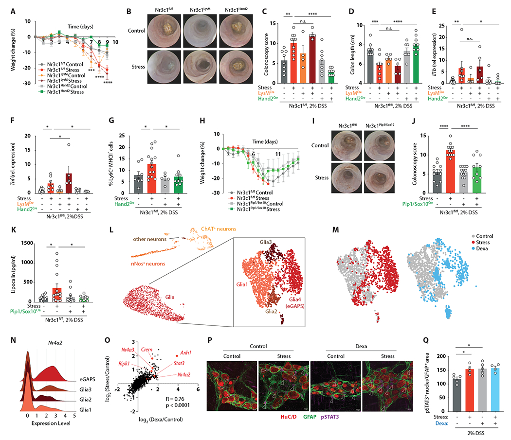
Mental health profoundly impacts inflammatory responses in the body. This is particularly apparent in inflammatory bowel disease (IBD), in which psychological stress is associated with exacerbated disease flares. Here, we discover a critical role for the enteric nervous system (ENS) in mediating the aggravating effect of chronic stress on intestinal inflammation. We find that chronically elevated levels of glucocorticoids drive the generation of an inflammatory subset of enteric glia that promotes monocyte- and TNF-mediated inflammation via CSF1. Additionally, glucocorticoids cause transcriptional immaturity in enteric neurons, acetylcholine deficiency, and dysmotility via TGF-β2. We verify the connection between the psychological state, intestinal inflammation, and dysmotility in three cohorts of IBD patients. Together, these findings offer a mechanistic explanation for the impact of the brain on peripheral inflammation, define the ENS as a relay between psychological stress and gut inflammation, and suggest that stress management could serve as a valuable component of IBD care.

Keywords: IBD; enteric glia; enteric nervous system; enteric neurons; glucocorticoids; monocytes; neuro-immune interactions; psychological stress.


Conflict of interest statement

Declaration of interests E.J.W. is an advisor for Danger Bio, Janssen, New Limit, Marengo, Pluto Immunotherapeutics Related Sciences, Rubius Therapeutics, Santa Ana Bio, Synthekine, and Surface Oncology. E.J.W. is a founder of and holds stock in Surface Oncology, Danger Bio, and Arsenal Biosciences.

Figures

Figure 1.
Figure 1.. Psychological stress exacerbates intestinal inflammation via colonic myeloid cells.
(A) Schematic of experimental stress-colitis paradigm. (B-E) Colitis severity readouts. (F) UMAP embedding of 23,696 colon leukocytes after 7 days of stress, including subclustering of myeloid cell populations. (G) Pseudotime trajectory analysis comparing control vs. stressed myeloid cells as well as TNF expression in Mono1 and Mac1 subclusters. (H) Flow cytometry of CD45+ Ly6GCD11b+ live colonic leukocytes. Bar charts depict relative abundances of cells in the respective quadrants (Q1-Q3). (I-K) Absolute numbers of Ly6C+ MHCII cells (I), Ly6C+ MHCII+ (J), Ly6C MHCII+ cells (K) from panel H. (L-N) Colitis readouts in CCR2-DTR mice. (O-Q) Colitis readouts in anti-TNF-treated mice. See also Figures S1–S4.
Figure 2.
Figure 2.. Psychological stress impacts intestinal inflammation through glucocorticoid signaling.
(A) Serum corticosterone after 1, 7, and 14 days of stress. (B-I) Colitis readouts in RU-486-treated (B-D) and dexamethasone-treated (E-I) stressed mice. (J) Flow cytometry of CD45+ Ly6G CD11b+ live colonic leukocytes after 7 days of dexamethasone treatment. Bar charts depict relative abundances of cells in the respective quadrants (Q1-Q3). (K-M) Absolute abundance of cells in Q1-Q3 from panel J. (N-Q) Colitis readouts of anti-TNF treated mice receiving dexamethasone. See also Figure S5.
Figure 3.
Figure 3.. The ENS relays the detrimental effect of glucocorticoids on intestinal inflammation.
(A-F) Colitis readouts (A-D) and relative gene expression levels of Il1b (E) and Tnf (F) in Nr3c1fl/fl, Nr3c1Hand2, and Nr3c1LysM mice. (G) Relative abundance of Ly6C+ MHCII cells determined by flow cytometry after an adjusted experimental paradigm (only 4 days of 2% DSS). (H-K) Colitis readouts in Nr3c1fl/fl and Nr3c1Sox10/Plp1 mice. (L) UMAP embedding of 9,858 enteric nuclei from Sox10Cre-INTACT mice after 7 days of stress with subclustering of glia cells. (M) UMAP embedding showing the distribution of cells from control (grey), stress (red) and dexamethasone-treated (blue) mice. (N) Ridgeline plot showing relative transcript levels of Nr4a2 in individual glia cell clusters. (O) Spearman correlation of the avg log2FC expression of significantly regulated genes in stress compared to control and dexamethasone-treated compared to control nuclei. (P and Q) Whole-mount IF staining for HuC/D, GFAP and pSTAT3 (arrows indicate pSTAT3+ nuclei) (P), and quantification of pSTAT3+ enteric glia cell nuclei normalized to the GFAP+ area (Q) after 7 days of stress or dexamethasone treatment. See also Figure S6 and S7.
Figure 4.
Figure 4.. Psychological stress drives colitis exacerbation via inflammatory enteric glia and CSF1.
(A-D) Colonoscopy readouts of stressed iDTRSox10 and iDTRfl/fl mice (A and B) and of dexamethasone-treated iDTRPlp1 and iDTRfl/fl mice (C and D) (E-G) Colitis readouts of BRAINSPAReDT-treated stressed mice. (H and I) Flow cytometry of CD45+ Ly6G live colonic leukocytes (H) and relative abundance of CD11b+Ly6Chi cells (I) from iDTRplp1 or iDTRfl/fl mice after 7 days of stress. (J) Circos plot showing significant cell-cell interactions determined by NicheNet. (K) Relative expression of Csf1 in enteric glia of control and stress mice determined by snRNA-seq. (L) CSF1 protein concentration in the colonic muscularis layer after 7 days of stress. (M) Percentage of colonic lymphocytes (live CD45+ CD19+/CD3+/CD5+) or Ly6Chi monocytes (live CD45+ Ly6G, CD11b+ Ly6Chi) expressing CSF1R as determined by flow cytometry. (N) Relative abundance of Ly6Chi MHCII (as a percentage of living CD45+ Ly6G CD11b+) cells in anti-CSF1 treated mice. (O-Q) Colitis readouts of mice treated with anti-CSF1. (R) Schematic of proposed pathway linking psychological stress to exacerbated colitis. See also Figures S6 and S7.
Figure 5.
Figure 5.. Psychological stress causes dysmotility via transcriptional immaturity in enteric neurons.
(A and B) Intestinal transit time after 7 days of stress (A) or dexamethasone treatment (B). (C) UMAP embedding of enteric nuclei isolated from colon of control and stressed Sox10Cre-INTACT mice after 7 days of stress. (D) Heatmap of differentially expressed genes across pseudotime. (E) Diffusion pseudotime after 7 days of stress. (F) Relative Nestin expression in bulk RNA-sequencing of total colon after 7 days of stress. (G and H) Whole-mount IF staining for HuC/D and Nestin (G) and quantification of Nestin+ cells (H). (I and J) Immunoblot analysis of colonic muscularis for Nestin (I) and quantification of relative Nestin protein levels (J) after 7 days of stress. (K-N) Whole-mount IF staining and quantifications of HuC/D, nNOS and ChAT (GFP) (K-L) and acetylcholine (ACh) concentrations in total colon tissue (N) after 7 days of stress. (O) Relative abundance of cells with pseudotime<0.3 after 7 days of dexamethasone treatment (determined by snRNA-seq). (P-S) Whole-mount IF staining and quantifications of HuC/D, nNOS and ChAT (GFP) (P-R), and acetylcholine (ACh) concentrations in total colon tissue (S) after 7 days of dexamethasone treatment. (T) Intestinal transit time after 7 days of stress in nicotine-treated mice. See also Figure S8.
Figure 6.
Figure 6.. Enteric neuronal immaturity contributes to stress-induced dysmotility via TGFβ2.
(A-D) Whole-mount IF staining and quantification of HuC/D, nNOS and Nestin in Nr3c1fl/fl and Nr3c1Hand2 mice exposed to stress for 7 days followed by 4 days of 2% DSS in drinking water. (E) Volcano plot of differentially expressed genes in neurons in low vs. high diffusion pseudotime. (F) Normalized Tgfb2 expression in the ENS nuclei of control, stress, and dexamethasone-treated mice (determined by snRNA-seq). (G and H) Relative Tgfb2 expression in colon determined by bulk mRNA sequencing (G) and TGFβ2 protein concentrations in colon muscularis layer (H) after 7 days of stress. (I-K) Whole-mount IF staining and quantifications of HuC/D, nNOS and Nestin in anti-TGFβ treated mice after a shortened experimental paradigm (only 4 days of 2% DSS). (L) Intestinal transit time after 7 days of stress and anti-TGFβ treatment. (M-O) Colitis readouts of anti-TGFβ treated mice. (P) Schematic of the proposed mechanism linking psychological stress to enteric dysmotility. See also Figure S8.
Figure 7.
Figure 7.. Psychological stress exacerbates IBD in humans.
(A) Workflow of data analysis from the UK Biobank. (B) Cumulative risk (adjusted for age, sex, BMI) for developing IBD in control vs. stressed participants. (C) Serum levels of C-reactive protein in control vs. stressed IBD patients. (D) Kaplan-Meier survival curve of control and stressed IBD patients with adjusted hazard ratio for age, sex, BMI. (E and F) Leukocyte (E) and monocyte (F) counts in the blood of control vs. stressed participants with IBD. (G) Dissatisfaction with bowel habits (scale max 10) in control vs. stressed IBD patients. (H) Presence of obstipation (in %) in control vs. stressed IBD patients. (I) Schematic of real-world telehealth study evaluating the effect of psychosocial stress on IBD severity. (J) Spearman’s correlation of questionnaire-based stress score and IBD activity score (MIAH). (K) Schematic of prospective study evaluating the effect of emotional stress on IBD severity. (L) Spearman’s correlation of perceived stress score and colonoscopy score (0-3). (M-R) Spearman’s correlation of perceived stress score with expression of the indicated genes. See also Figure S9 and Table S1.

Comment in

References

    1. Haykin H, and Rolls A (2021). The neuroimmune response during stress: A physiological perspective. Immunity 54, 1933–1947. 10.1016/j.immuni.2021.08.023. - DOI - PMC - PubMed
    1. Liu YZ, Wang YX, and Jiang CL (2017). Inflammation: The common pathway of stress-related diseases. Frontiers in Human Neuroscience. 10.3389/fnhum.2017.00316. - DOI - PMC - PubMed
    1. Qing H, Desrouleaux R, Israni-Winger K, Mineur YS, Fogelman N, Zhang C, Rashed S, Palm NW, Sinha R, Picciotto MR, et al. (2020). Origin and Function of Stress-Induced IL-6 in Murine Models. Cell 182, 372–387.e14. 10.1016/jcel12020.05.054. - DOI - PMC - PubMed
    1. Chang JT (2020). Pathophysiology of Inflammatory Bowel Diseases. The New England journal of medicine 383, 2652–2664. 10.1056/NEJMra2002697. - DOI - PubMed
    1. Sgambato D, Miranda A, Ranaldo R, Federico A, and Romano M (2017). The Role of Stress in Inflammatory Bowel Diseases. Current Pharmaceutical Design. 10.2174/1381612823666170228123357. - DOI - PubMed

Publication types

Substances

LinkOut - more resources