Review
. 2023 Jul;22(7):100584.
doi: 10.1016/j.mcpro.2023.100584. Epub 2023 May 24.

In-Depth Characterization of Apoptosis N-Terminome Reveals a Link Between Caspase-3 Cleavage and Posttranslational N-Terminal Acetylation

Affiliations
Review

In-Depth Characterization of Apoptosis N-Terminome Reveals a Link Between Caspase-3 Cleavage and Posttranslational N-Terminal Acetylation

Rawad Hanna et al. Mol Cell Proteomics. 2023 Jul.

Abstract

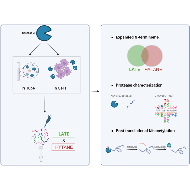
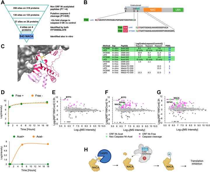
The N termini of proteins contain information about their biochemical properties and functions. These N termini can be processed by proteases and can undergo other co- or posttranslational modifications. We have developed LATE (LysN Amino Terminal Enrichment), a method that uses selective chemical derivatization of α-amines to isolate the N-terminal peptides, in order to improve N-terminome identification in conjunction with other enrichment strategies. We applied LATE alongside another N-terminomic method to study caspase-3-mediated proteolysis both in vitro and during apoptosis in cells. This has enabled us to identify many unreported caspase-3 cleavages, some of which cannot be identified by other methods. Moreover, we have found direct evidence that neo-N-termini generated by caspase-3 cleavage can be further modified by Nt-acetylation. Some of these neo-Nt-acetylation events occur in the early phase of the apoptotic process and may have a role in translation inhibition. This has provided a comprehensive overview of the caspase-3 degradome and has uncovered previously unrecognized cross talk between posttranslational Nt-acetylation and caspase proteolytic pathways.

Keywords: LysN; N-terminal acetylation; N-terminomics; caspase-3; degradomics; peptidyl-Lys metalloendopeptidase.


Conflict of interest statement

Conflict of interest The authors declare that they have no conflicts of interest with the contents of this article.

Figures

None
Graphical abstract
Fig. 1
Fig. 1
LysN Amino Terminal Enrichment (LATE) workflow for improved N-terminome coverage.A, in silico analysis of the “Identifiable” and “Not identifiable” putative caspase-3 DEVD↓X in the human proteome following digestion by trypsin with ArgC-like specificity (red) and LysN (green). B, LATE workflow for the comparison of two conditions. Extracted proteins of each sample are digested with LysN, and the resulting peptides undergo N-terminal-specific isotopic dimethylation. The samples are then mixed and subject to hydrophobic tagging of lysines, which allows for their removal and for the enrichment of the N-terminal peptides. The drawing was created with BioRender.com. C, LATE N-terminal dimethylation labeling specificity following LysN digestion. Over 95% of peptides’ N-terminal amines were labeled by dimethylation or naturally blocked by acetylation while less than 5% of the lysine side-chain amines were labeled. D, peptide-spectrum match (PSM) number of LysN internal peptides (green) versus the PSM number of ORF (burgundy) and neo N-terminal peptides (blue) before (PreLATE) and after LATE. E, comparison of human proteins with free (left) and acetylated (right) N-terminal peptides identified by HYTANE (red) and LATE (green). HYTANE, hydrophobic tagging-assisted N-termini enrichment; LATE, LysN amino terminal enrichment.
Fig. 2
Fig. 2
N-terminomics of caspase-3 cleavages in vitro.A, in vitro experimental scheme. The drawing was created with BioRender.com. B, ranked ratios of dimethylated peptides showing the accumulation trends of peptides with high Log2 (caspase-3/control) abundance ratio (>1 as marked by the yellow dotted line) in LATE and HYTANE. C, Venn diagram of peptides with Log2 (caspase-3/control) >1 at each time point in LATE and in HYTANE. D, Venn diagram of the unique cleavage sites after D or E that were identified only in the caspase-3-treated samples with a caspase-3/control ratio ≥2 in at least one time point. “Reported/not reported” is based on a comparison with published data. E, sequence logo of all putative cleavage sites that were identified only in the caspase-3-treated samples with a caspase-3/control ratio ≥2, in comparison with all caspase-3 cleavage found in MEROPS. F, distribution of all putative cleavage sites that were identified only in the caspase-3-treated samples with a caspase-3/control ratio ≥2 based on the amino acid at the P1 position of their cleavage motif. G, the residue distance distribution of the nearest arginine to the identified peptide sequence in LATE (green) and HYTANE (red) experiments. HYTANE, hydrophobic tagging-assisted N-termini enrichment; LATE, LysN amino terminal enrichment.
Fig. 3
Fig. 3
N-terminomics of caspase-3 cleavages in cells.A, cell-based experimental scheme to study caspase-3-mediated cleavages in HCT116 following treatment with ABT-199. The drawing was created with BioRender.com. B, number of N-terminal peptides identifications following HYTANE and LATE enrichments. C, sequence logo of neo-N-terminal peptides cleavage motifs by HYTANE (top) and LATE (bottom). D, Venn diagram of the combined number of neo cleavage motifs identified by HYTANE (red) and LATE (green). E, the number of putative caspase cleavage motifs (with P1 = D or E) out of the total neo-N-terminal cleavage motifs that were identified. The categorization of reported/not reported is based on a comparison with published data (TopFind, etc.). F, the number of putative caspase cleavage motifs (with P1 = D or E) that had a high caspase-3/control abundance ratio (Log2 ≥ 1) out of the total neo-N-terminal cleavages identified. G, novel caspase-3 substrates and their cleavage sites. The cleavage site (caspase-3/control) abundance ratio is indicated by color. HYTANE, hydrophobic tagging-assisted N-termini enrichment; LATE, LysN amino terminal enrichment.
Fig. 4
Fig. 4
Acetylation of both ORF and neo N-terminal peptides.A, the ORF N-terminal peptides identified in HCT116 cells overexpressing caspase-3 and their control were categorized based on the presence of initiation methionine (marked as first Met) or its removal (marked as second position) and the presence of Nt-acetylation (“Nt-Acet”; in shades of blue) or its absence (“Free”; in shades of pink). B, the abundance ratio distribution of the ORF N-terminal peptides in cells overexpressing caspase-3 and their control, based on their N-terminal state (free/acetylated) and the presence of Asp or Glu residue in their sequence. Box limits indicate the 25th and 75th percentiles; whiskers extend 1.5 times the interquartile range from the 25th and 75th percentiles; crosses represent sample means; data points are plotted as open circles. n = 128, 808, 133, 620 sample points. C, the abundance ratio distribution of the 122 ORF N-terminal peptides that were identified both as Nt-acetylated and with free N terminus and contain Asp or Glu in their sequence. Center lines show the medians; box limits indicate the 25th and 75th percentiles; whiskers extend 1.5 times the interquartile range from the 25th and 75th percentiles; data points are plotted as open circles. D, posttranslational neo-Nt-acetylation sites that were identified at N termini generated following known proteolytic processing of precursor proteins as indicated in UniProt. The blue diamonds represent identification in biological repeats or time points. HYTANE, hydrophobic tagging-assisted N-termini enrichment; LATE, LysN amino terminal enrichment.
Fig. 5
Fig. 5
Neo-Nt-acetylation of nascent polypeptide–associated complex A (NACA) of ORF and neo-N-terminal peptides.A, winnowing of acetylated neo-N-terminal peptides identified in HCT116 cells. B, the N-terminal peptides of nascent polypeptide–associated complex subunit alpha (NACA) identified in HCT116 cells. Cleaved peptide sequences and modifications are shown on the top and the peptide-spectrum match (PSM) numbers in the different experiments are on the bottom. C, the position of NACA cleavage (in yellow and light blue) by caspase-3, before Ser43, lead to the formation of a neo-Nt-acetylated form of NACA. The structural model is based on the structure of the ribosome-nascent chain containing an endoplasmic reticulum signal sequence in a complex with the nascent polypeptide–associated complex (Protein Data Bank: 7QWR). The ribosome and RNA are in gray. The full NACA (in dark pink) and BTF3 (in light pink) structures are based on AlphaFold (86) prediction and were aligned on top of the original partial structures. D, time-course of in vitro NACA cleavage and Nt-acetylation. The upper panel shows NACA cleavage at Ser43 by caspase-3 with (green) or without (orange) lysate dialysis before caspase-3 addition. The lower panel shows NACA neo-acetylation at Ser43 after caspase-3 cleavage with (green) or without (orange) lysate dialysis before caspase-3 addition. E, F and G, time-course N-terminomics of HCT116 cells treated with ABT-199 or DMSO (control). Log2 abundance ratios for individual peptides are plotted on the y-axis, and the corresponding total peptide mass spectrometry (MS) intensities are shown on the x-axis. The circle colors correspond to the peptide type: ORF Nt-Acetylated (light gray), ORF peptides with Nt-free (dark gray), caspase-generated neo-Nt peptides (pink), and neo-Nt-acetylated peptides (light green). H, the suggested order of events for NACA neo-Nt-acetylation following caspase-3 processing. HYTANE, hydrophobic tagging-assisted N-termini enrichment; LATE, LysN amino terminal enrichment.

References

    1. Lopez-Otin C., Overall C.M. Protease degradomics: a new challenge for proteomics. Nat. Rev. Mol. Cell Biol. 2002;3:509–519. - PubMed
    1. López-Otín C., Bond J.S. Proteases: multifunctional enzymes in life and disease. J. Biol. Chem. 2008;283:30433–30437. - PMC - PubMed
    1. Schlage P., Egli F.E., auf dem Keller U. Methods in Molecular Biology. Humana Press; New York, NY: 2017. pp. 185–198. - PubMed
    1. Marino G., Eckhard U., Overall C.M. Protein termini and their modifications revealed by positional proteomics. ACS Chem. Biol. 2015;10:1754–1764. - PubMed
    1. Sukharev S.A., Pleshakova O.V., Sadovnikov V.B. Role of proteases in activation of apoptosis. Cell Death Differ. 1997;46:457–462. - PubMed

Publication types