. 2023 Jun 28;43(26):4926-4940.
doi: 10.1523/JNEUROSCI.1988-22.2023. Epub 2023 May 26.

Neuronally Derived Soluble Abeta Evokes Cell-Wide Astrocytic Calcium Dysregulation in Absence of Amyloid Plaques in Vivo

Affiliations

Neuronally Derived Soluble Abeta Evokes Cell-Wide Astrocytic Calcium Dysregulation in Absence of Amyloid Plaques in Vivo

Patricia Kelly et al. J Neurosci. .

Abstract

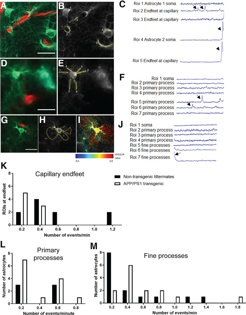
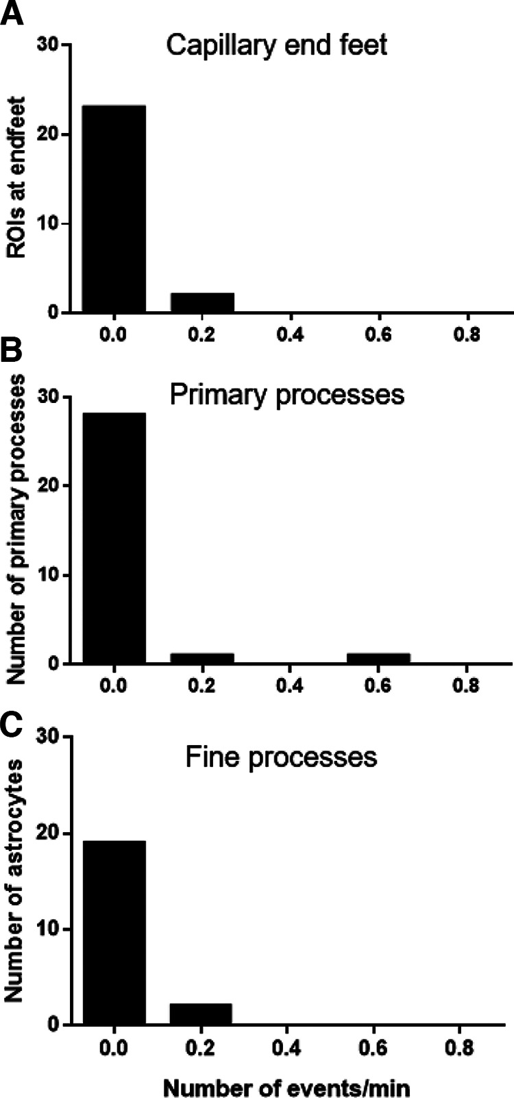
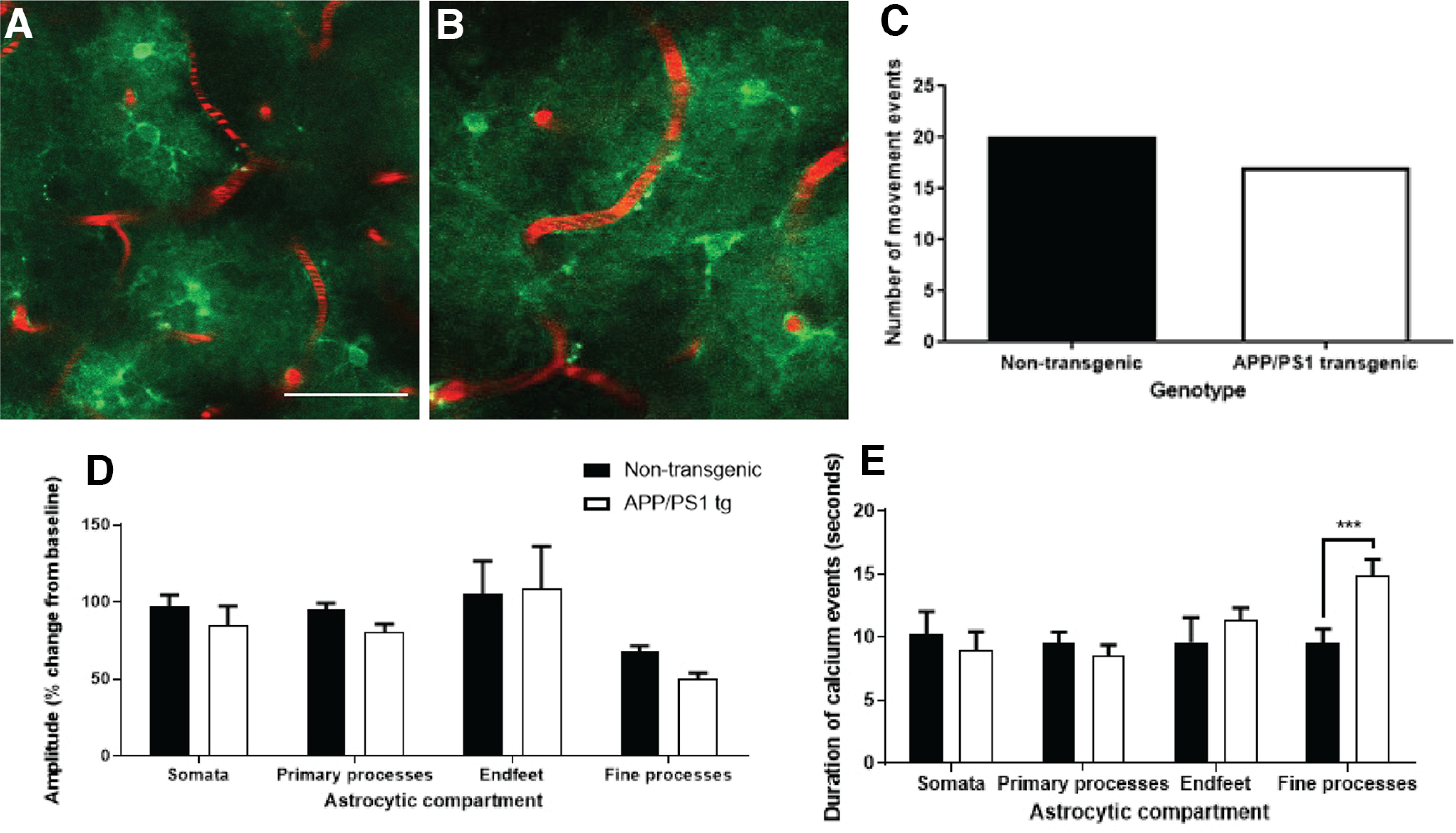
The key pathologic entities driving the destruction of synaptic function and integrity during the evolution of Alzheimer's disease (AD) remain elusive. Astrocytes are structurally and functionally integrated within synaptic and vascular circuitry and use calcium-based physiology to modulate basal synaptic transmission, vascular dynamics, and neurovascular coupling, which are central to AD pathogenesis. We used high-resolution multiphoton imaging to quantify all endogenous calcium signaling arising spontaneously throughout astrocytic somata, primary processes, fine processes, and capillary endfeet in the brain of awake APP/PS1 transgenic mice (11 male and 6 female mice). Endogenous calcium signaling within capillary endfeet, while surprisingly as active as astrocytic fine processes, was reduced ∼50% in the brain of awake APP/PS1 mice. Cortical astrocytes, in the presence of amyloid plaques in awake APP/PS1 mice, had a cell-wide increase in intracellular calcium associated with an increased frequency, amplitude, and duration of spontaneous calcium signaling. The cell-wide astrocytic calcium dysregulation was not directly related to distance to amyloid plaques. We could re-create the cell-wide intracellular calcium dysregulation in the absence of amyloid plaques following acute exposure to neuronally derived soluble Abeta from Tg2576 transgenic mice, in the living brain of male C57/Bl6 mice. Our findings highlight a role for astrocytic calcium pathophysiology in soluble-Abeta mediated neurodegenerative processes in AD. Additionally, therapeutic strategies aiming to protect astrocytic calcium physiology from soluble Abeta-mediated toxicity may need to pharmacologically enhance calcium signaling within the hypoactive capillary endfeet while reducing the hyperactivity of spontaneous calcium signaling throughout the rest of the astrocyte.SIGNIFICANCE STATEMENT Astrocytic calcium signaling is functionally involved in central pathologic processes of Alzheimer's disease. We quantified endogenous calcium signaling arising spontaneously in the brain of awake APP/PS1 mice, as general anesthesia suppressed astrocytic calcium signaling. Cell-wide astrocytic calcium dysregulation was not related to distance to amyloid plaques but mediated in part by neuronally derived soluble Abeta, supporting a role for astrocytes in soluble-Abeta mediated neurodegeneration. Spontaneous calcium signaling is largely compartmentalized and capillary endfeet were as active as fine processes but hypoactive in the presence of amyloid plaques, while the rest of the astrocyte became hyperactive. The cell-wide calcium pathophysiology in astrocytes may require a combination therapeutic strategy for hypoactive endfeet and astrocytic hyperactivity.

Keywords: Abeta; Alzheimer's disease; astrocytes; calcium signaling; in vivo imaging.


Figures

Figure 1.
Figure 1.
Cell-wide astrocytic intracellular calcium dysregulation unrelated to distance to amyloid plaques in awake brain of APP/PS1 mice. A, Maximum projection images showing network of gfa2.yc3.6-expressing astrocytes (green) and cerebral vasculature (red) in the brain of an awake nontransgenic mouse. Corresponding pseudo-colored image showing the YFP/CFP ratio of each astrocyte as low (blue) to high (red; B) and age-matched APP/PS1 transgenic mouse, with gfa2.yc3.6-expressing astrocytes, vasculature, and methoxy-labeled amyloid plaque (C, cyan; corresponding pseudo-colored image, D). Astrocytic intracellular calcium (YFP/CFP ratios) is significantly elevated within astrocytic somata (E; p = 0.0109; parametric t test), astrocytic primary processes (F; p = 0.0172; Mann–Whitney test), and capillary endfeet (G; p = 0.0116; parametric t test) in awake brain of APP/PS1 mice (n = 10) compared with awake brain of nontransgenic mice (n = 9). The cell-wide intracellular calcium dysregulation within astrocytic somata (p = 0.6845; H), primary processes (p = 0.4987; I), and capillary endfeet (p = 0.3724; J) was not influenced by distance to amyloid plaques in the brain of awake APP/PS1 mice. Data are mean ± SEM. Scale bar, 25 µm.
Figure 2.
Figure 2.
gfa2.yc3.6-expressing astrocytes colocalize with SR-101-positive astrocytes within living mouse brain and immunohistochemical astrocyte markers glutamine synthetase and GFAP in mouse brain sections. At the top, AAV2/5.gfa2.yc3.6-expressing astrocytes (A) and Sulforhodamine 101 (SR-101) fluorescently labeled astrocytes (B) colocalized within the living cortex of a 6-month-oldÜ57BL/6J mouse in vivo (C). At the bottom, Immunohistochemical staining of gfa2.yc3.6-expressing astrocytes within 8-month-old C57BL/6J mouse brain sections shows colocalization with either glutamine synthetase-labeled astrocytes (D–F) or GFAP-labeled astrocytes (G–I), but not NeuN-positive neurons (A–C). Scale bars: 25 μm, 50 μm.
Figure 3.
Figure 3.
Neuronally derived soluble Abeta evokes a cell-wide calcium overload in cortical astrocytes, in the absence of amyloid plaques in the brain of young C57BL/6J mice, during anesthesia by isoflurane. A, Schematic representation of the experimental procedure to determine the effects of neuronally derived soluble Abeta on astroglial resting calcium in the healthy mouse brain in vivo. B, Representative in vivo images of gfa2.yc3.6 expressing astrocytes (green) and fluorescently labeled cerebral blood vessels (red) in brain of young C57BL/6J. Astrocytes within white box with dotted lines shown at higher magnification for baseline in vivo imaging and at 1 h following topical application of ntgCM, TgCM, or Aβ-immunodepleted transgenic conditioned media. C, Histograms of astrocytic calcium frequency distribution (YFP/CFP ratio) for the three conditions before (basal) and after application of CM (1 h). The percentage of astrocytic cellular compartments exceeding the threshold for calcium overload (black dotted line; 2 SDs greater than the YFP/CFP mean determined for the baseline) is noted on the graphs. D, Relative increase in astroglial [Ca2+] (ΔR/R0) after conditioned media application for each volume acquired. E, Percentage of calcium overload (YFP/CFP ratios .2 SDs above mean of YFP/CFP ratios for nontransgenic mice) before and after application of conditioned media for every mouse analyzed. Acute exposure to TgCM significantly increased calcium overload within astrocytic somata (from 2.2% to 11.8%, p < 0.001), primary processes (from 1.9% to 7.7%, p < 0.05), and astrocytic endfeet (from 1.8% to 8.0%, p < 0.05). Error bars indicate mean ± SEM. ***p < 0.001. **p < 0.005. n = 5-7 mice per group. Scale bars: 20 µm; insets, 5 µm.
Figure 4.
Figure 4.
Chronic astrocytic calcium overload in awake brain of APP/PS1 mice. Representative pseudo-colored in vivo image showing high YFP/CFP ratio (red) in gfa2.yc3.6-expressing astrocyte (white arrowhead) adjacent to a methoxy-X04-positive amyloid plaque (blue; A) longitudinally imaged in brain of awake APP/PS1 mice each month (B,C) revealing chronic elevated calcium within robust cortical astrocytes.
Figure 5.
Figure 5.
Neuronally derived soluble Abeta increases astrocytic intracellular calcium in vitro. A, Representative images of astrocytic cell line WJE before and following acute 1 h exposure to either ntgCM cultured from neurons from nontransgenic mice or TgCM cultured from neurons from Tg2576 transgenic mice. B, Quantification of YFP/CFP ratios for every WJE cell before and after either nTgCM or TgCM. C, Averaged YFP/CFP ratios before (basal) and after acute exposure to nTgCM or TgCM per experiment. ****p < 0.0001; **p < 0.005; two-tailed t test. n = 3 or 4 experiments. Scale bar, 50 μm.
Figure 6.
Figure 6.
Astrocytic intracellular “resting” calcium not compartmentalized throughout somata and primary processes in somatosensory cortex of awake mice. Astrocytic intracellular calcium within somata is strongly correlated with the average intracellular calcium of all corresponding primary processes of the cell in the somatosensory cortex of awake 12- to 17-month-old nontransgenic mice (96 astrocytes; n = 2 mice; r = 0.7905, p < 0.0001; Pearson correlation coefficient two-tailed) and awake APP/PS1 mice (249 astrocytes; n = 5 mice; r = 0.7110, p < 0.0001; nonparametric Spearman correlation two-tailed).
Figure 7.
Figure 7.
Compartmentalized spontaneous calcium signaling hypoactive within capillary endfeet alongside astrocytic hyperactivity in awake APP/PS1 mice. A–I, Representative time-lapse in vivo images of gfa2.yc3.6-expressing astrocytes (green), vasculature (red), and amyloid plaques (cyan) acquired within the somatosensory cortex of awake mice. Aligned image, using custom-written MATLAB script, demonstrating positioning of manually drawn ROIs (B,E,H) and corresponding spatiotemporal dynamics of all endogenous compartmentalized calcium signaling events occurring within ROIs during 300 s time-lapse indicated by black arrows (C,F,J). Spontaneous calcium wave traveling along capillary endfeet at ∼33 μm/s and spreading into adjacent astrocytic somata and across the cortex in concentric wave (C, black arrowheads; Movie 1). Qualitative pseudo-colored time-lapse in vivo image showing high YFP/CFP ratio occurring solely within astrocytic fine processes (I) and corresponding calcium signaling event indicated by black arrow (J). K, Compartmentalized calcium signaling is very active within capillary endfeet (0.2-1.2 events/min) in awake nontransgenic mice but hypoactive within the brain of awake APP/PS1 mice (0.2-0.4 events/min). L, Endogenous calcium signaling compartmentalized within astrocytic primary processes is less active than capillary endfeet in awake nontransgenic mice (0.2-0.6 events/min) and awake APP/PS1 mice (0.2-0.8 events/min). M, Spontaneous calcium signaling compartmentalized within astrocytic fine processes is very active within nontransgenic mice (0.2-1.4 events/min) and APP/PS1 mice (0.2-1.8 events/min). Scale bar, 20 µm.
Figure 8.
Figure 8.
Amyloid plaque environment did not appear to influence cell-wide quiescence in cortical astrocytes in brain of awake APP/PS1 mice. Representative in vivo time-lapse image of gfa2.yc3.6-expressing astrocyte within awake brain of nontransgenic mouse (A) and manually drawn ROIs for quantification of spontaneous calcium signaling throughout entire cell (B). C, An example of cell-wide quiescence with no spontaneous calcium events occurring in any of the cellular compartments during 300 s time-lapse. D, In vivo time-lapse image showing quiescent cortical astrocyte in awake brain of APP/PS1 mouse, with no spontaneous calcium events occurring within any of the manually drawn ROIs (E) over 300 s time-lapse (F). G, Approximately 37% of cortical astrocytes within the somatosensory cortex of nontransgenic mice show cell-wide quiescence over 300 s compared with 41% cortical astrocytes with cell-wide quiescence in awake APP/PS1 mice. Scale bar, 20 µm.
Figure 9.
Figure 9.
Use of isoflurane (2%) during in vivo multiphoton imaging largely suppressed all types of compartmentalized spontaneous calcium events within cortical astrocytic processes in the living brain of 4- to 6-month-old C57BL/6J mice. A, Isoflurane use silenced compartmentalized spontaneous calcium events within 23 of 25 ROIs at endfeet at 19 cerebral capillaries in the brain of C57BL/6 mice. Compartmentalized spontaneous calcium events were also silenced within 28 of 30 primary processes (B) and fine processes of 21 cortical astrocytes (C) in the anesthetized brain of C57BL/6 mice (n = 6 mice).
Figure 10.
Figure 10.
Mouse movement-associated global calcium signaling occurring with hyperactivity within astrocytic fine processes in awake APP/PS1 mice. Representative in vivo time-lapse images of gfa2.yc3.6-expressing astrocytes (green) and vasculature (red) within the cortical network of awake nontransgenic mice (A) and APP/PS1 transgenic mice (B). C, Mice were habituated to being awake during in vivo multiphoton imaging, and movement events associated with global astrocytic calcium signaling were similar between groups. D, Global astrocytic calcium signaling associated with mouse movement occurred with similar amplitude throughout all astrocytic cellular compartments in awake APP/PS1 mice and nontransgenic mice (p = 0.6983; two-way ANOVA). E, Astrocytic calcium signaling within fine processes was hyperactive during movement-induced calcium signaling in awake APP/PS1 mice compared with nontransgenic mice (p = 0.0010; two-way ANOVA). Data are mean ± SEM. Scale bar, 50 µm.
Figure 11.
Figure 11.
Astrocytic calcium signaling involving multiple cellular compartments can emanate from any cellular compartment and appear hyperactive in awake APP/PS1 mice. A, Representative in vivo time-lapse images showing gfa2.yc3.6-expressing astrocyte (green) and fluorescently labeled vasculature (red) in awake mouse brain. Manually drawn ROIs (B) and corresponding calcium signaling for each ROI show spontaneous calcium signaling occurring simultaneously within somata and primary processes (C). D, The number of multicompartmental events occurring per minute (0.2-0.6 events/s) was not different between groups. E, There was significantly greater amplitude of calcium signaling within astrocytic somata in APP/PS1 mice compared with nontransgenic mice (p = 0.0066; two-way ANOVA). F, Duration of calcium signaling was significantly greater within astrocytic fine processes in APP/PS1 mice compared with nontransgenic mice (p = 0.0089; two-way ANOVA). Multicompartmental calcium signaling events in astrocytes can first appear within any cellular compartment (G) but most commonly occur within primary processes in awake APP/PS1 mouse brain (H). Data are mean ± SEM. Scale bar, 20 µm.
Figure 12.
Figure 12.
Hyperactive spontaneous intracellular calcium activity within astrocytic processes and concomitant hypoactivity within astrocytic endfeet in the amyloid plaque environment in the brain of awake APP/PS1 mice. Proportional segments of 98 spontaneous intracellular calcium events occurring throughout astrocytes within brain of awake WT mice (A) and 111 spontaneous intracellular calcium events occurring throughout astrocytes within brain of awake APP/PS1 mice (B). Compartmentalized calcium activity within capillary endfeet (28%) as active as fine processes (33%) in brain of awake nontransgenic mice (A) but reduced to 10% in brain of APP/PS1 mice while spontaneous calcium signaling within primary processes show hyperactivity (B).
Figure 13.
Figure 13.
Quantification of spontaneous “multicellular” calcium events occurring throughout cortical astrocytes within the brain of awake mice. A, Two cortical astrocytes adjacent to fluorescently labeled small blood vessels (red) within the awake brain of nontransgenic mouse. B, Aligned time-lapse image with manually drawn ROIs. C, YFP/CFP ratio traces, for 700 frames and 300 s, showing two spontaneous multicellular calcium events that occupy all cellular compartments of both astrocytes in A, B. D–G, Spatiotemporal tracking of spontaneous multicellular events, with sequence of event propagation highlighted by numerical order, showing heterogeneity between first and second occurrence of a multicellular event during 300 s of time-lapse acquisition within the awake mouse brain. H, Spontaneous multicellular calcium events occur with greater amplitude within astrocytic somata and primary processes in cortical astrocytes within the awake brain of APP/PS1 mice compared with nontransgenic mice (p = 0.01; two-way ANOVA). Data are mean ± SEM. Scale bar, 20 µm.

References

    1. Abdul HM, Sama MA, Furman JL, Mathis DM, Beckett TL, Weidner AM, Patel ES, Baig I, Murphy MP, LeVine H 3rd, Kraner SD, Norris CM (2009) Cognitive decline in Alzheimer's disease is associated with selective changes in calcineurin/NFAT signaling. J Neurosci 29:12957–12969. 10.1523/JNEUROSCI.1064-09.2009 - DOI - PMC - PubMed
    1. Arbel-Ornath M, Hudry E, Boivin JR, Hashimoto T, Takeda S, Kuchibhotla KV, Hou S, Lattarulo CR, Belcher AM, Shakerdge N, Trujillo PB, Muzikansky A, Betensky RA, Hyman BT, Bacskai BJ (2017) Soluble oligomeric amyloid-β induces calcium dyshomeostasis that precedes synapse loss in the living mouse brain. Mol Neurodegener 12:27. 10.1186/s13024-017-0169-9 - DOI - PMC - PubMed
    1. Attwell D, Buchan AM, Charpak S, Lauritzen M, MacVicar BA, Newman EA (2010) Glial and neuronal control of brain blood flow. Nature 468:232–243. 10.1038/nature09613 - DOI - PMC - PubMed
    1. Bazargani N, Attwell D (2016) Astrocyte calcium signaling: the third wave. Nat Neurosci 19:182–189. 10.1038/nn.4201 - DOI - PubMed
    1. Biesecker KR, Srienc AI, Shimoda AM, Agarwal A, Bergles DE, Kofuji P, Newman EA (2016) Glial cell calcium signaling mediates capillary regulation of blood flow in the retina. J Neurosci 36:9435–9445. 10.1523/JNEUROSCI.1782-16.2016 - DOI - PMC - PubMed

Publication types