. 2023 Jul 5;43(27):5030-5044.
doi: 10.1523/JNEUROSCI.2170-22.2023. Epub 2023 May 26.

Cortically Evoked Movement in Humans Reflects History of Prior Executions, Not Plan for Upcoming Movement

Affiliations

Cortically Evoked Movement in Humans Reflects History of Prior Executions, Not Plan for Upcoming Movement

Abdelbaset Suleiman et al. J Neurosci. .

Abstract

Human motor behavior involves planning and execution of actions, some more frequently. Manipulating probability distribution of a movement through intensive direction-specific repetition causes physiological bias toward that direction, which can be cortically evoked by transcranial magnetic stimulation (TMS). However, because evoked movement has not been used to distinguish movement execution and plan histories to date, it is unclear whether the bias is because of frequently executed movements or recent planning of movement. Here, in a cohort of 40 participants (22 female), we separately manipulate the recent history of movement plans and execution and probe the resulting effects on physiological biases using TMS and on the default plan for goal-directed actions using a timed-response task. Baseline physiological biases shared similar low-level kinematic properties (direction) to a default plan for upcoming movement. However, manipulation of recent execution history via repetitions toward a specific direction significantly affected physiological biases, but not plan-based goal-directed movement. To further determine whether physiological biases reflect ongoing motor planning, we biased plan history by increasing the likelihood of a specific target location and found a significant effect on the default plan for goal-directed movements. However, TMS-evoked movement during preparation did not become biased toward the most frequent plan. This suggests that physiological biases may either provide a readout of the default state of primary motor cortex population activity in the movement-related space, but not ongoing neural activation in the planning-related space, or that practice induces sensitization of neurons involved in the practiced movement, calling into question the relevance of cortically evoked physiological biases to voluntary movements.SIGNIFICANCE STATEMENT Human motor performance depends not only on ability to make movements relevant to the environment/body's current state, but also on recent action history. One emerging approach to study recent movement history effects on the brain is via physiological biases in cortically-evoked involuntary movements. However, because prior movement execution and plan histories were indistinguishable to date, to what extent physiological biases are due to pure execution-dependent history, or to prior planning of the most probable action, remains unclear. Here, we show that physiological biases are profoundly affected by recent movement execution history, but not ongoing movement planning. Evoked movement, therefore, provides a readout of the default state within the movement space, but not of ongoing activation related to voluntary movement planning.

Keywords: TMS; evoked movement; history of movement; motor planning; primary motor cortex.


Figures

Figure 1.
Figure 1.
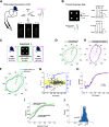
Experimental setup, protocol, and results of experiment 1. A, Schematic overview of the TMS protocol. We delivered TMS over the motor cortex to elicit thumb movements and measured physiological bias as the evoked movement with maximum probability. Top right, Dashed lines represent multiple evoked thumb movements. Bottom, The forearm and the index to the pinky fingers were immobilized and strapped to the armrest using a Velcro strap; only the thumb was free to move. The angles indicated the direction within a two-dimensional movement space (shown on the screen) and not anatomic range of motion. B, Timed-response task. Participants controlled the cursor presented on a monitor by voluntarily moving their thumb. In regular trials, participants initiated their movement synchronously with the onset of the fourth tone toward a target (top). In catch trials, no target ever appeared, but participants were still required to plan and move to any direction with the onset of the fourth tone (bottom). RT indicates effective reaction time. C, Protocol of experiment 1. Participants performed a block of TMS (65 pulses), followed by a block of goal-directed timed-response tasks (150 trials), and last, a block of TMS (65 pulses). The second TMS block was conducted to make sure that the relatively short block of voluntary movements (150 trials) did not lead to significant physiological biases. D, von Mises probability distribution of physiological biases (i.e., TMS-evoked movements; magenta) and plan-based biases (green). E, Distributions of physiological biases in TMS blocks 1 and 2, indicating that short practice of voluntary movement was not sufficient to induce significant physiological changes. Data in D, E are from all participants pooled together. F, The distribution of movement directions in the timed-response trials occurring any time before the target appeared and up to 50 ms immediately after the target was displayed (dark green) were comparable to the directions in the catch trails (light green). G, Example of data during the timed-response condition pooled across participants in experiment 1 (target 2). Gray points indicate direction error (degree) as a function of RT for individual trials; blue line shows the moving average of the probability that a movement is successful for a given RT, which corresponds to the speed-accuracy trade-off. Yellow area indicates the range of where a given movement was considered successful. H, Illustration of maximum likelihood model fit (red line) to empirical speed-accuracy trade-off data (blue line). Arrow indicates the estimated parameters α0 that defined the participant's lower limit accuracy. I, Speed-accuracy trade-off for movements near the plan-based direction revealed that this movement involved the default plan. This was reflected by the increased accuracy at short RTs for targets near the plan-based bias but not for the other targets. J, Fitting the speed-accuracy function revealed significant difference of the parameter α0 (which reflects participant's lower limit accuracy) between near plan-based bias (near-PBB) target and the other targets (far-PBB).
Figure 2.
Figure 2.
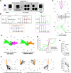
Movement history modulated physiological biases, not plan-based bias of voluntary movements. A, Protocol of experiment 2. B, von Mises probability distribution of physiological biases (left) and plan-based biases (right) before (dashed outlines) and after (solid outlines) changing probability distribution of prior movements through repetition toward a novel direction. Data from an individual participant. C, Histogram of change (after-before) in direction of physiological biases (magenta) and plan-based biases (green). D, Cumulative distribution function of differences in directions for physiological biases, plan-based biases, and null hypothesis of normal distribution (black dashed line). E, The distribution of movement directions in the timed-response trials any time before the target appeared and up to 50 ms immediately after the target was displayed (dark green) were comparable to the plan-based directions in the catch trials (light green). F, Speed-accuracy trade-off for movements near the plan-based direction revealed that this movement involved the default plan. This was reflected by the increased accuracy at short RTs for targets near the plan-based biases, but not for the other targets. G, Fitting the speed-accuracy function revealed significant difference of the parameter α0 (which reflects participant's lower limit accuracy) between near plan-based bias (near-PBB) target and the other targets (far-PBB).
Figure 3.
Figure 3.
Movement-related physiological changes were distinct from ongoing planning-related processing. A, Protocol of experiment 3. B, Trial schedule of each trial type. During Go/No-Go trials, the target appeared 750 ms before the cue (Target turned green in Go trials, whereas in No-Go trials the target turned red.). In some trials, after presenting the target, a single TMS pulse was delivered 150 ms before the Go/No-Go cue. This allowed sufficient time to prepare the desired movement. During the postplan repetition block, we implemented the timed-response task that included some catch trials where target never appeared. In addition, in some of these catch trials, a single TMS pulse was delivered 150 ms before the fourth tone. C, von Mises probability distribution of physiological biases (top) before (dashed magenta), during (solid orange) and after (solid magenta) the plan repetition block. von Mises probability distribution of plan-based biases (bottom) before (dashed green) and after (solid green) the plan repetition block. Data from an individual participant. D, Histogram of change in direction of physiological biases (plan repetition-before, orange; after-before, magenta), and plan-based biases (green). E, Cumulative distribution function of differences in directions for physiological biases, plan-based biases, and null hypothesis of normal distribution. F, Thumb movements (black lines) during the Go trials and TMS-evoked thumb movement during ongoing planning of the desired movement (orange lines). Each circle represents the distribution of movement for each of the four targets during the plan repetition block. Data suggest robust and invariant physiological biases regardless of the plan. G, Reaction time (seconds) for movements in the Go trials toward the most frequent target (black) and the other targets (gray). sec, Seconds. Early and late represent the first and last 10 trials of the repetition block, respectively. Average across participants (top) and individual data are shown (bottom). Data show mean ± SE, *p < 0.05, **p < 0.01, ****p < 0.0001.
Figure 4.
Figure 4.
Proposed dynamic of neural activity in the motor cortex underlies physiological and plan-based biases. Recent execution history alters the default state in the movement-related space toward the repeated direction, with little effect on ongoing activity of the preparatory space. Stimulation of M1 may thus reflect a readout of the cortical activity where movements located close to the default state may be more likely to be elicited by TMS. On the other hand, manipulating plan-based expectation changed the default preparatory state.

Similar articles

Cited by

References

    1. Afshar A, Santhanam G, Yu BM, Ryu SI, Sahani M, Shenoy KV (2011) Single-trial neural correlates of arm movement preparation. Neuron 71:555–564. 10.1016/j.neuron.2011.05.047 - DOI - PMC - PubMed
    1. Ames KC, Ryu SI, Shenoy KV (2014) Neural dynamics of reaching following incorrect or absent motor preparation. Neuron 81:438–451. 10.1016/j.neuron.2013.11.003 - DOI - PMC - PubMed
    1. Bestmann S (2012) Functional modulation of primary motor cortex during action selection. In: Cortical connectivity (Chen R, Rothwell J, eds), pp 183–205. Berlin: Springer.
    1. Bestmann S, Duque J (2016) Transcranial magnetic stimulation: decomposing the processes underlying action preparation. Neuroscientist 22:392–405. 10.1177/1073858415592594 - DOI - PubMed
    1. Bestmann S, Krakauer JW (2015) The uses and interpretations of the motor-evoked potential for understanding behaviour. Exp Brain Res 233:679–689. 10.1007/s00221-014-4183-7 - DOI - PubMed

Publication types

LinkOut - more resources