Review
. 2023 Jul;24(7):416-430.
doi: 10.1038/s41583-023-00701-0. Epub 2023 May 26.

The impact of the human thalamus on brain-wide information processing

Affiliations
Review

The impact of the human thalamus on brain-wide information processing

James M Shine et al. Nat Rev Neurosci. 2023 Jul.

Abstract

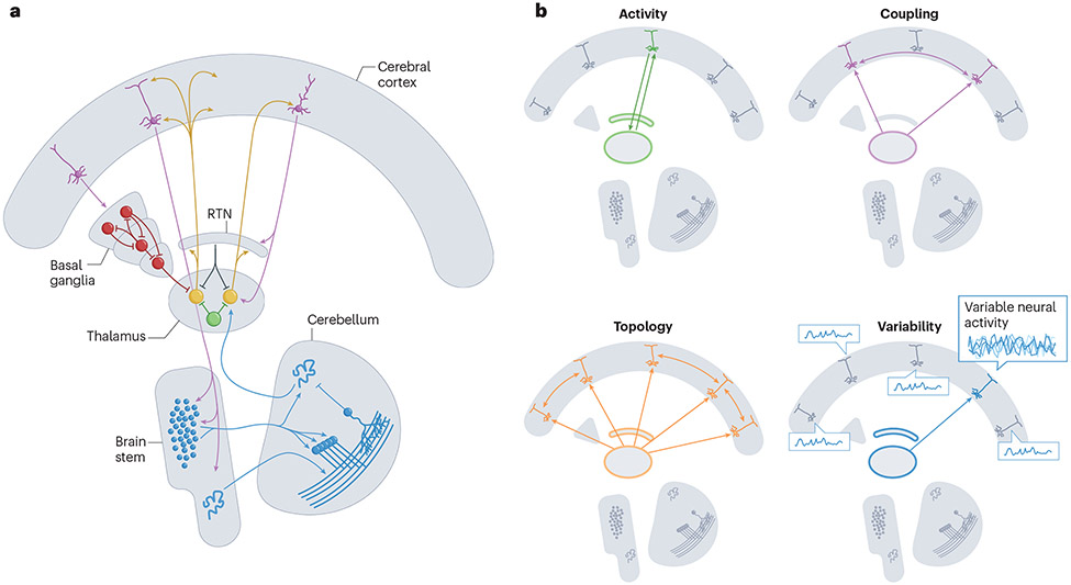
The thalamus is a small, bilateral structure in the diencephalon that integrates signals from many areas of the CNS. This critical anatomical position allows the thalamus to influence whole-brain activity and adaptive behaviour. However, traditional research paradigms have struggled to attribute specific functions to the thalamus, and it has remained understudied in the human neuroimaging literature. Recent advances in analytical techniques and increased accessibility to large, high-quality data sets have brought forth a series of studies and findings that (re-)establish the thalamus as a core region of interest in human cognitive neuroscience, a field that otherwise remains cortico-centric. In this Perspective, we argue that using whole-brain neuroimaging approaches to investigate the thalamus and its interaction with the rest of the brain is key for understanding systems-level control of information processing. To this end, we highlight the role of the thalamus in shaping a range of functional signatures, including evoked activity, interregional connectivity, network topology and neuronal variability, both at rest and during the performance of cognitive tasks.


Figures

Figure 1.
Figure 1.. The functional neuroanatomy of the thalamus.
A) The thalamus is embedded in a distributed neural architecture: different populations of neurons in the thalamus (orange and brown) are interconnected with pyramidal neurons in the cerebral cortex (purple), inhibitory input from the basal ganglia (red), excitatory input from the cerebellum (blue) and local inhibition, both from the reticular nucleus (RTN; grey) and GABAergic interneurons (INs; green). B) The thalamus has a crucial role in at least four partially overlapping canonical functions: promoting focused cortical activity (green), facilitating inter-regional coupling (purple), supporting changes in network topology (orange) and dimensionality (that is, the higher-order structure of the interactions between neurons), and enabling and modulating temporal neuronal variability (blue).
Figure 2.
Figure 2.. Functional parcellation, connector hub properties and variability profile of the thalamus.
A) Thalamic regions associate with cortical functional networks that are putatively involved in cognitive control related functions , namely the cingulo-opercular network (CON), the frontoparietal network (FPN), the saliency network (SAL) and the dorsal attention network (DAN). B) Top: The medial, mediodorsal, and posterior portions of the thalamus exhibit particularly strong connector hub properties, which are indicated by their higher scores on the participation coefficient (PC) axis. The colour bar depicts the PC scale, which is a measure of connector hubness and ranges from 0 to 1 (low to high). Bottom: Most of the thalamus exhibits connector hub properties that are stronger than those of cortical regions. C) The mean blood oxygen level-dependent (BOLD) signal in three different thalamic groups — the medial (pink), posterior (blue) and anterolateral (orange) — is strongly aligned with the parametric effect of cognitive load on the low-dimensional distributed activity — coloured bars designate significant parametric effects (p < 0.05 from non-parametric permutation testing) and the dotted black (tPC*1) line depicts mean low-dimensional trajectories. This indicates a low-dimensional relationship between activity in the thalamus and activity in the cerebral cortex. D) Toy example of three functional interaction scenarios between the prefrontal cortex and the thalamus. Here, the expectation is that with increasing functional connection (that is, going from 1 (green) to 2 (blue) to 3 (black) connected nodes; left panel), moment-to-moment brain signal variability expressed by the thalamus should increase in kind (middle panel). Accordingly, higher temporal variability in the thalamus may reflect higher functional integration among connected regions. E) Heightened BOLD variability in thalamus was the strongest signature of lower principal component analysis (PCA) dimensionality (that is, higher moment-to-moment functional integration between brain regions). F) Thalamic regions projecting to the prefrontal cortex were particularly sensitive to joint longitudinal changes between BOLD signal variability, functional integration and cognition; individuals who maintained moment-to-moment thalamic variability also maintained functional integration and cognition over 2.5 years. Part a adapted with permission from REF.. Part b adapted with permission from REF.. Part c adapted with permission from REF.. Parts d and e adapted with permission from REF.. Part f adapted with permission from REF..
Figure 3.
Figure 3.. Arousal shapes thalamic activity and thalamocortical dynamics.
A) Schematic of individual nuclei of the thalamus imaged with 7T functional MRI (fMRI), locked to arousal state transitions. A temporal sequence of activity emerges across thalamic nuclei, preceding the moment of transition to higher arousal states. Shading shows 95% confidence interval for activity timing in each thalamic nucleus, locked to arousal. B) A spatial template of key fMRI predictors of arousal shows thalamic correlations with arousal state. The colour intensity shows the strength of each region’s correlation with behavioural arousal measured via eye closures; thalamus shows strong positive correlations. C) Inducing sedation with dexmedetomidine, a drug that induces decreased noradrenergic neuromodulatory tone, causes decreased thalamic glucose metabolism and connectivity. AV, anteroventral nucleus; CM, centromedian nucleus; dmPFC, dorsomedial prefrontal cortex; LGN, lateral geniculate nucleus; MD, mediodorsal nucleus; PCC, posterior cingulate cortex; PUL, pulvinar nucleus; RSC, retrosplenial cortex; VA, ventral anterior nucleus; VLA, ventral lateral anterior nucleus; VLP, ventral lateral posterior nucleus; vmPFC, ventromedial prefrontal cortex; VPL, ventral posterolateral nucleus. Part a adapted with permission from REF.. Part b adapted with permission from REF.. Part c adapted with permission from REF..
Figure 4.
Figure 4.. The functional repertoire of the thalamus serves decision-making under parametric uncertainty.
A) In the multi-attribute attention task, participants were first cued with a set of 1-4 potentially task relevant stimulus features (any set of color, direction, size, or luminance). They were then shown a stimulus that contains all four features, and were subsequently asked to make a decision about one feature. Participants underwent fMRI and EEG during the task, in separate sessions. Results indicated that B) participants with larger parametric increases in thalamic BOLD (especially in antero-medial nuclei that project to frontoparietal cortical targets) were more likely to C) upregulate EEG-based ‘excitability’ (reduced alpha and increased gamma, expressed by a spectral power modulation component (SPMC); flatter 1/f spectral slopes; higher sample entropy (SampEn), exhibit a higher drift rate and drift rate modulation, and show heightened arousal (first derivative of pupil responses). D) Those who expressed higher uncertainty-related modulation of behavioural drift rate expressed higher thalamic modulation particularly during stimulus presentation (yellow shading). Figure adapted with permission from REF..

Similar articles

Cited by

References

    1. Luo TZ & Maunsell JHR Attention can be subdivided into neurobiological components corresponding to distinct behavioral effects. Proc Natl Acad Sci USA 116, 26187–26194 (2019). - PMC - PubMed
    1. Bullmore E & Sporns O The economy of brain network organization. Nature Reviews Neuroscience 13, 336–349 (2012). - PubMed
    1. Buschman TJ & Miller EK Top-Down Versus Bottom-Up Control of Attention in the Prefrontal and Posterior Parietal Cortices. Science 315, 1860–1862 (2007). - PubMed
    1. Krienen FM, Yeo BTT & Buckner RL Reconfigurable task-dependent functional coupling modes cluster around a core functional architecture. Phil. Trans. R. Soc. B 369, 20130526 (2014). - PMC - PubMed
    1. Shine JM et al. The Dynamics of Functional Brain Networks: Integrated Network States during Cognitive Task Performance. Neuron 92, 544–554 (2016). - PMC - PubMed