. 2023 May 26;13(1):8551.
doi: 10.1038/s41598-023-35632-4.

Heterologous production of the D-cycloserine intermediate O-acetyl-L-serine in a human type II pulmonary cell model

Affiliations

Heterologous production of the D-cycloserine intermediate O-acetyl-L-serine in a human type II pulmonary cell model

Laurel Robbins et al. Sci Rep. .

Abstract

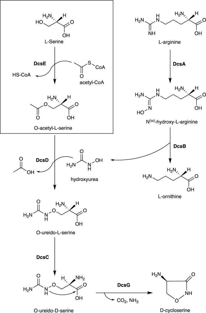
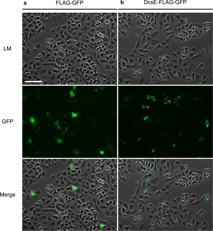
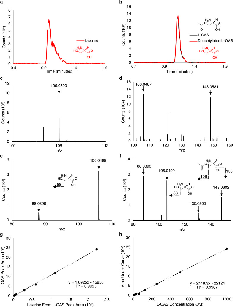
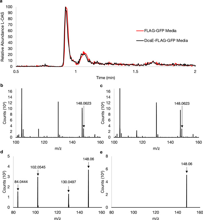
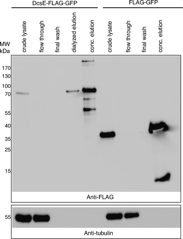
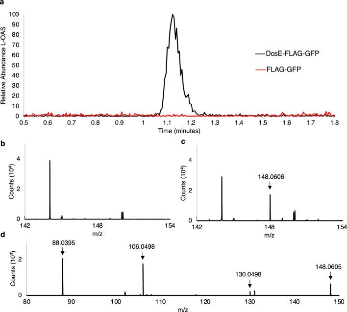
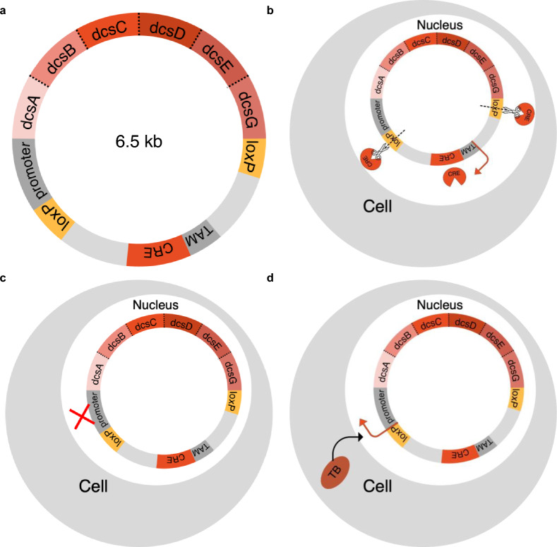
Tuberculosis (TB) is the second leading cause of death by a single infectious disease behind COVID-19. Despite a century of effort, the current TB vaccine does not effectively prevent pulmonary TB, promote herd immunity, or prevent transmission. Therefore, alternative approaches are needed. We seek to develop a cell therapy that produces an effective antibiotic in response to TB infection. D-cycloserine (D-CS) is a second-line antibiotic for TB that inhibits bacterial cell wall synthesis. We have determined D-CS to be the optimal candidate for anti-TB cell therapy due to its effectiveness against TB, relatively short biosynthetic pathway, and its low-resistance incidence. The first committed step towards D-CS synthesis is catalyzed by the L-serine-O-acetyltransferase (DcsE) which converts L-serine and acetyl-CoA to O-acetyl-L-serine (L-OAS). To test if the D-CS pathway could be an effective prophylaxis for TB, we endeavored to express functional DcsE in A549 cells as a human pulmonary model. We observed DcsE-FLAG-GFP expression using fluorescence microscopy. DcsE purified from A549 cells catalyzed the synthesis of L-OAS as observed by HPLC-MS. Therefore, human cells synthesize functional DcsE capable of converting L-serine and acetyl-CoA to L-OAS demonstrating the first step towards D-CS production in human cells.


Conflict of interest statement

The authors declare no competing interests.

Figures

Figure 1
Figure 1
Biosynthetic pathway for D-CS,. Boxed reaction catalyzed by DcsE. D-cycloserine can be produced biosynthetically using six enzymes, DcsA-G, (in bold) and biologically available reagents: L-serine, L-arginine, and acetyl-CoA.
Figure 2
Figure 2
A549 cells express FLAG-GFP and DcsE tagged with FLAG and GFP. (a) GFP in FLAG-GFP transfected A549 cells (negative control) indicates expression of GFP-FLAG and more diffused protein. (b) GFP in DcsE-FLAG-GFP transfected A549 cells indicates more punctate expression of DcsE-FLAG-GFP. Light microscopy (LM), Green Fluorescent Protein (GFP). Scale bar is 100 µm.
Figure 3
Figure 3
Retention times, MS, MSMS results, and standard curves for L-serine and L-OAS in reaction buffer using HPLC–MS. (a) HPLC chromatogram showing retention time vs counts for 500 µM L-serine (0.9 min). (b) HPLC chromatogram of a pure 125 µM L-OAS standard. L-OAS (black) and the fragmentation of L-OAS to L-serine (red) appear at the retention time of 1.1 min. (c) ESI-TOF positive mode peak for L-serine 106.05 m/z (molecular weight (MW) of L-serine 105.09). (d) ESI-TOF positive mode peak for L-OAS 148.0581 m/z (MW of L-OAS 147.13) and fragmented L-OAS to L-serine 106.0499 m/z. (e) Tandem MS for L-serine at 5 V shows a single fragment of 88.0396 m/z and intact L-serine at 106.0499 m/z. (f) Tandem MS for L-OAS at 5 V show fragments of 88.0396, 106.0499, 130.0500, and intact L-OAS at 148.0602 m/z. (g) Linear relationship between peak area of L-OAS from standard curve and the peak area of fragmented L-OAS to L-serine. (h) Standard curve of L-OAS (concentrations ranging from 1 mM to 7.8 µM). Inset chemical schematic depict fragmentation consistent with fragment ions observed.
Figure 4
Figure 4
No detection of L-OAS from the media of A549 cells transfected with DcsE-FLAG-GFP or FLAG-GFP. (a) Chromatogram of suspected L-OAS (148 m/z) detected from FLAG-GFP (red) and DcsE-FLAG-GFP (black) transfection media. (b, c) ESI-TOF peaks of 148.058 m/z representing suspected L-OAS at 1.14 min from FLAG-GFP (b) and DcsE-FLAG-GFP (c) transfection media. (d,e) Tandem MS of 148 m/z from (b) and (c), respectively, show fragments of 84.0444, 102.0545, 130.0497, and 148.06 m/z at 5 V. The expected 88.0396 and 106.05 fragment ions from an L-OAS standard were not detected.
Figure 5
Figure 5
DcsE-FLAG-GFP is purified and concentrated following FLAG immunoprecipitation. Western blot analysis of indicated fractions from purification of DcsE-FLAG-GFP (67 kDa) or FLAG-GFP (28 kDa) from transfected A549 cells. 7 µl of indicated fraction was loaded per lane. Membrane was first probed with anti-FLAG antibody and then stripped and reprobed using anti-tubulin as detailed in materials and methods. Full sized blots and ladder images are provided as supplementary data (Fig. S2).
Figure 6
Figure 6
Synthesis of D-CS intermediate, L-OAS, by heterologous purified DcsE-FLAG-GFP. Purified DcsE reacted with L-Serine and Acetyl-CoA was functional as determined by presence of enzyme product, L-OAS using HPLC–MS as analyzed by MassHunter. (a) HPLC chromatogram of L-OAS produced by DcsE-FLAG-GFP (in black) at 1.117 min. L-OAS was not detected from a reaction containing FLAG-GFP (in red). (b) FLAG-GFP containing samples produce no detectable peak of 148.06 m/z at 1.117 min suggesting no L-OAS production. (c) DcsE-FLAG-GFP containing samples produce ESI-TOF peak of 148.06 m/z at 1.117 min which corresponds to mass of L-OAS. (d) tandem MS of L-OAS produced by DcsE create fragments at 88.04, 106.05, and 131.03 m/z.
Figure 7
Figure 7
Proposed excisable D-CS plasmid for TB-inducible expression. (a) Map of proposed D-CS plasmid. Infection responsive element promoter (grey: InfRE), tamoxifen inducible promoter (grey: TAM), DcsABCDEG and CRE genes (reds), excisable loxP sites (gold), Mtb MycP1 cleavage sites SLKPASAGGG (dotted lines). (b) Cell with excised plasmid activated by tamoxifen which expresses CRE recombinase to cut at loxP sites. (c) Cell not expressing DcsABCDEG in the case of no TB infection. (d) Cell expressing DcsABCDEG in the case of TB infection.

References

    1. Koch, R. The etiology of tuberculosis. Berl. Klin. Wocbenschift10, 221–230 (1882).
    1. World Health Organization. Global tuberculosis report 2021. https://www.who.int/publications-detail-redirect/9789240037021 (2021).
    1. Principi, N. & Esposito, S. The present and future of tuberculosis vaccinations. Tuberculosis95, 6–13 (2015). - PubMed
    1. Tameris, M. et al. The candidate TB vaccine, MVA85A, induces highly durable Th1 responses. PLoS ONE9, e87340 (2014). - PMC - PubMed
    1. Smieja, M., Marchetti, C., Cook, D. & Smaill, F. M. Isoniazid for preventing tuberculosis in non‐HIV infected persons. Cochrane Database Syst. Rev.1999, CD001363 (1999). - PMC - PubMed

Publication types