Review
. 2023 May 26;30(1):33.
doi: 10.1186/s12929-023-00925-3.

The biology of SCUBE

Affiliations
Review

The biology of SCUBE

Yuh-Charn Lin et al. J Biomed Sci. .

Abstract

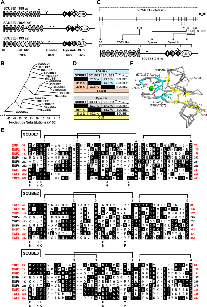
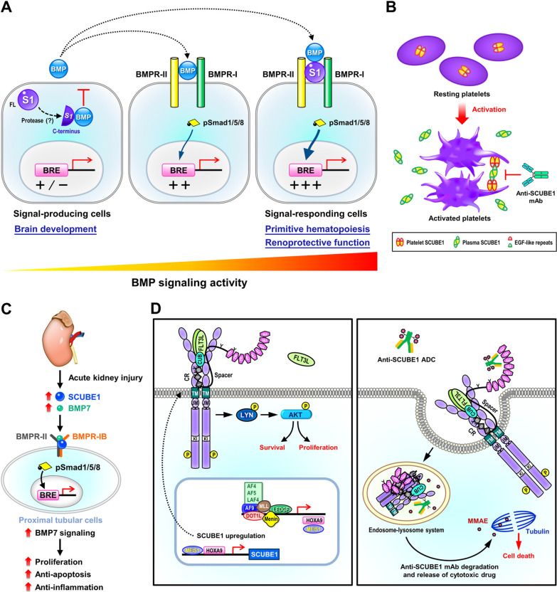
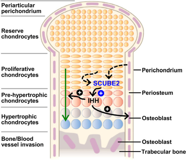
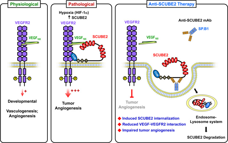
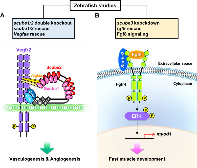
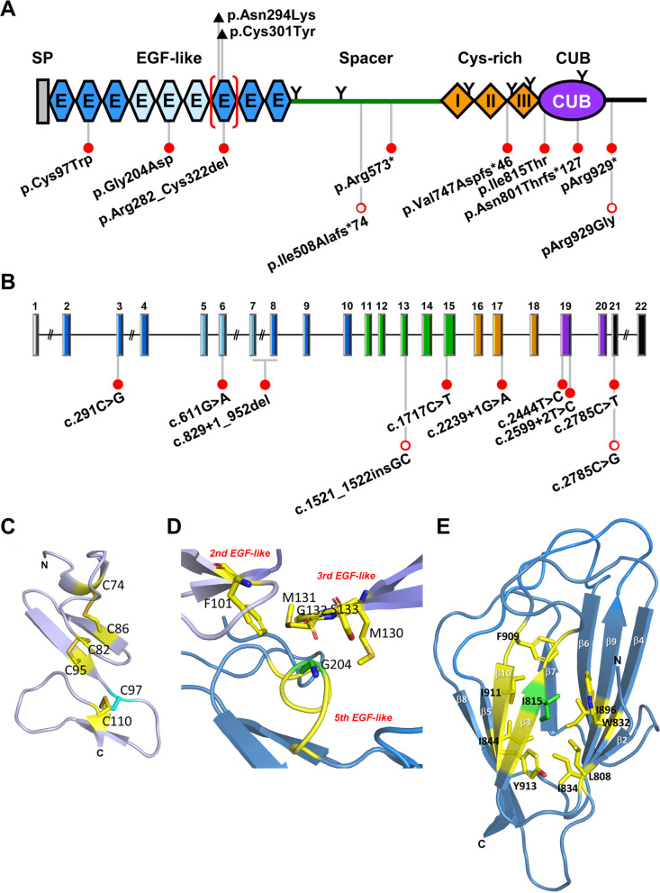
The SCUBE [Signal peptide-Complement C1r/C1s, Uegf, Bmp1 (CUB)-Epithelial growth factor domain-containing protein] family consists of three proteins in vertebrates, SCUBE1, 2 and 3, which are highly conserved in zebrafish, mice and humans. Each SCUBE gene encodes a polypeptide of approximately 1000 amino acids that is organized into five modular domains: (1) an N-terminal signal peptide sequence, (2) nine tandem epidermal growth factor (EGF)-like repeats, (3) a large spacer region, (4) three cysteine-rich (CR) motifs, and (5) a CUB domain at the C-terminus. Murine Scube genes are expressed individually or in combination during the development of various tissues, including those in the central nervous system and the axial skeleton. The cDNAs of human SCUBE orthologs were originally cloned from vascular endothelial cells, but SCUBE expression has also been found in platelets, mammary ductal epithelium and osteoblasts. Both soluble and membrane-associated SCUBEs have been shown to play important roles in physiology and pathology. For instance, upregulation of SCUBEs has been reported in acute myeloid leukemia, breast cancer and lung cancer. In addition, soluble SCUBE1 is released from activated platelets and can be used as a clinical biomarker for acute coronary syndrome and ischemic stroke. Soluble SCUBE2 enhances distal signaling by facilitating the secretion of dual-lipidated hedgehog from nearby ligand-producing cells in a paracrine manner. Interestingly, the spacer regions and CR motifs can increase or enable SCUBE binding to cell surfaces via electrostatic or glycan-lectin interactions. As such, membrane-associated SCUBEs can function as coreceptors that enhance the signaling activity of various serine/threonine kinase or tyrosine kinase receptors. For example, membrane-associated SCUBE3 functions as a coreceptor that promotes signaling in bone morphogenesis. In humans, SCUBE3 mutations are linked to abnormalities in growth and differentiation of both bones and teeth. In addition to studies on human SCUBE function, experimental results from genetically modified mouse models have yielded important insights in the field of systems biology. In this review, we highlight novel molecular discoveries and critical directions for future research on SCUBE proteins in the context of cancer, skeletal disease and cardiovascular disease.

Keywords: Biomarker; Coreceptor; Endothelial cells; SCUBE; Signal transduction.


Conflict of interest statement

The authors declare no competing interests.

Figures

Fig. 1
Fig. 1
Protein domains, phylogenetic analysis, genomic organization, sequence alignment, and sequence identity of SCUBE protein family. A Graphic illustration of the domain structure of human SCUBE1, 2 and 3 (see Table 1). SP, signal peptide sequence; E, EGF-like domain (grey shade indicated the calcium-binding EGF module); Cys-rich, cysteine-rich repeats; CUB, the CUB domain. “Y” marks the potential N-linked glycosylated sites of each SCUBE protein. Protein domain sequence identity shared among human SCUBE members is calculated at the bottom; the highest homology was found in the CUB domain (83%), followed by the EGF-like (73%) and Cys-rich (66%) domains. The spacer region appears to have the lowest homology (34%) and may be associated with the unique functions of each SCUBE member. B Phylogenetic tree of the SCUBE family. Similarity of human (h), mouse (m), zebrafish (z), eagle (e), lizard (l), or frog (f) SCUBE protein sequences was analyzed by using the Lasegene MEGALIGN program (ClustalW algorithm). The length of each pair of branches represents the phylogenic distance between sequence pairs. Below the tree is a scale indicating the number of “Nucleotide Substitutions” for both DNA and protein sequences. C Genomic structure of human SCUBE1. SCUBE1 gene consists of 22 exons spanning about 140 kb on chromosome 22. The exon–intron boundaries are well preserved among the SCUBE gene members, which suggests possible gene duplication during evolution. Of note, SCUBE genomic organizations derived from zebrafish, mouse and human genomes (http://genome.ucsc.edu) follow a modular arrangement, with the signal peptide sequence, each epidermal growth factor (EGF)-like domain, and Cys-rich motif, each encoded by a single exon. In addition, the spacer region is encoded by five exons and the CUB is encoded by two exons. D Amino acid sequence identity of SCUBE family. Data in the upper right (blue shading) represent the sequence identity in EGF-like domain, and data on the bottom left (orange shading) represent the sequence identity in spacer region between SCUBE proteins (upper panel). Data on the upper right (gray shading) present the sequence identity in Cys-rich domain, and data on the bottom left (yellow shading) present the sequence identity in CUB domain between SCUBE proteins (lower panel). E Sequence alignment, sequence identity, and structural comparison of EGF-like repeats of human SCUBE proteins. Protein sequences of SCUBE1, SCUBE2 and SCUBE3 used for alignment were derived from NCBI Reference Sequence Database, as listed in Table 1. Symbols use the one-letter code for amino acids. A dash indicates a gap. The conserved cysteines are numbered 1 to 6, and the predicted disulfide bonds form as follows: C1-C3, C2-C4, and C5-C6. Amino acids indicated below highlight the canonical calcium binding (cb) consensus sequence (D/N)-X-(D/N)-E/Q-Xm (D/N*) Xn (Y/F), where m and n are variable and the asterisk indicates potential β-hydroxylation [17]. Of note, two triplets of cbEGF1-3 or cbEGF7-9 modules (amino acid residue boundaries marked in red fonts) are well conserved in all SCUBE members. F Structural comparison of SCUBE3 second and seventh cbEGF-like domains with human NOTCH1 fifth cbEGF-like domain. 3D models of the second and seventh EGF-like domains of human SCUBE3 obtained from AlphaFold2 (AF-Q8IX30-F1-model_v3) [187] were superimposed on the fifth cbEGF-like domain of human NOTCH1 (PDB 5FM9) [221]. These EGF-like domains contain a short calcium binding motif sequence signature: (D/N)-X-(D/N)-E/Q-Xm-(D/N*)-Xn-(Y/F), where m and n are variable and the asterisk indicates potential β-hydroxylation [17]. This canonical calcium coordination module is found near at the N-terminus. The residues coordinating calcium ion (in green) are shown as sticks, and the coordination as pink dotted lines. The five calcium-binding motif residues indicated in E are presented as cyan sticks; Asn and Phe/Try between C3 and C4 of these EGF-like domains are consistent with the substrate sequence pattern of Asp/Asn-β-hydroxylase [222, 223]. The three paired cysteines (C1:C3, C2:C4, and C5:C6) are presented as yellow sticks. Atoms are colored, with oxygen in red, nitrogen in blue and sulfur in gold. This figure was produced using PyMOL (Schrödinger, LLC. The PyMOL molecular graphics system, version 1.8, https://pymol.org). Nt, NH2 terminus; Ct, COOH terminus
Fig. 2
Fig. 2
Sequence alignment of the spacer regions, Cys-rich domains, and CUB domains in the human SCUBE family. The margins of the spacer region and the CUB domain are derived from PFAM analysis and are marked by arrows. Amino acids residues that are identical in at least 2 protein sequences are highlighted in black. Potential N-linked glycosylation sites shared among the SCUBE proteins are denoted with a filled red circle. The minimal furin cleavage sites (RXXR), which are present in SCUBE1 and SCUBE3 but not SCUBE2, are boxed
Fig. 3
Fig. 3
Pathological involvement of SCUBE1. A SCUBE1 modulates BMP signaling activity in a context-dependent manner. When expressed in “signal-producing” cells, the C-terminal CR and CUB domains are released by an as yet undetermined proteinase, allowing them to bind and inhibit the secretion of mature BMP into the culture medium. SCUBE1 therefore acts as an antagonist for long-range BMP signaling activity during brain development [20]. In contrast, when SCUBE1 is expressed in “signal-responding” cells, it forms a complex with BMP ligand and its receptors. SCUBE1 thereby acts as a BMP coreceptor to augment BMP signaling activity critical for protecting against kidney ischemia–reperfusion (I/R) injury or for primitive hematopoiesis [8, 49]. B Proposed model for adhesive function of SCUBE1 in platelet aggregation and thrombus formation. Under normal conditions, plasma SCUBE1 is expressed at low levels primarily by endothelial cells and platelets. In addition, SCUBE1 is stored in the α-granules of resting platelets. Upon pathological stimulation, activated platelets secrete large amounts of SCUBE1 into the circulation and high levels of SCUBE1 are also found on the platelet surface. The surface SCUBE1 on nearby activated platelets is trans-homophilically crosslinked by soluble SCUBE1, acting through its sticky EGF-like repeats to promote platelet agglutination and stabilize platelet plugs. Targeting the adhesive modules of SCUBE1 with a specific monoclonal antibody might be a potentially useful anti-thrombotic strategy [72, 86]. C SCUBE1-promoted BMP signaling protects against kidney ischemia/reperfusion (I/R) injury. I/R-induced SCUBE1 protein in proximal tubular epithelial cells serves as a BMP coreceptor to enable renoprotective BMP7 signaling, which stimulates epithelial repair and regeneration through proliferative, anti-apoptotic, and anti-inflammatory effects [49]. D Potential immunotherapy strategy and the pathological function of surface SCUBE1 in MLL-r leukemias. Left: SCUBE1 is an immediate downstream target of the HOXA9/MEIS1 transcriptional regulatory complex, which is activated by MLL-fusion proteins like MLL-AF9 and is crucial for sustaining leukemic transition. Surface SCUBE1 functions as a FLT3 coreceptor in MLL-r leukemias, facilitating the interaction between FLT3 ligand and FLT3. This action increases downstream LYN-AKT activation (tyrosine phosphorylation) to enhance leukemic cell proliferation and survival, thereby promoting leukemogenesis. Right: An anti-SCUBE1 ADC conjugated to an antimitotic agent (MMAE) leads to significant cell killing specifically in MLL-AF9 leukemias. Thus, surface expression of SCUBE1 on MLL-r leukemia cells may be useful as a target for immunotherapy. ADC antibody–drug conjugate; MMAE monomethyl auristatin E. MLL-r, mixed-lineage leukemia gene-rearranged; BMP bone morphogenetic protein 1
Fig. 4
Fig. 4
SCUBE2 is essential for controlling IHH signaling during endochondral ossification. SCUBE2 protein is expressed in peri-chondrial cells and may be secreted (dashed line with arrow). This secreted SCUBE2 could modulate IHH signals derived from pre-hypertrophic chondrocytes (solid line with arrow) by promoting the release and mobilization of IHH, which drives differentiation into osteoblasts during endochondral bone formation. Green arrow indicates the differentiation direction toward hypertrophic chondrocytes within endochondral bone. IHH Indian hedgehog
Fig. 5
Fig. 5
Physiological and pathological roles of endothelial SCUBE2 in modulating VEGF signaling during angiogenesis and potential immunotherapy approach. VEGF-stimulated VEGFR2 signaling is essential for angiogenesis and vasculogenesis under physiological conditions, such as during embryonic development (left panel). However, under pathological circumstances (e.g., intratumor hypoxia), hypoxia-inducible HIF-1 upregulates SCUBE2. The SCUBE2 protein localizes on the tumor EC cell surface, where it functions as a coreceptor with VEGFR2 to facilitate VEGF binding and enhance its downstream signaling. Thus, SCUBE2 promotes VEGF-induced tumor angiogenesis [23] (middle panel). SCUBE2 is internalized into the endosome-lysosomal protein degradation pathway when SP.B1 mAb attaches to EC-surface SCUBE2. This internalization decreases the VEGF-VEGFR2 association and prevents tumor angiogenesis (right panel). EC, endothelial cell. VEGF vascular endothelial growth factor; VEGFR2 VEGF receptor 2
Fig. 6
Fig. 6
Functional studies of SCUBE family using the zebrafish model. A Scube1 and Scube2 from zebrafish endothelial cells work together to support Vegfa signaling during embryonic vascularization. Endothelial Scube1 and Scube2 form a complex under physiological conditions, such as during the development of embryonic vessels. This complex may promote Vegf-induced Vegfr2 phosphorylation and its downstream signaling for proper vasculogenesis and angiogenesis [130]. B Zebrafish Scube3 acts as an Fgf coreceptor during fast muscle fiber development. We utilized a combination of molecular, biochemical, and genetic methods to reveal a new biological function for SCUBE3 as a key regulator of fast muscle precursors. SCUBE3 potentially acts as a coreceptor at the plasma membrane to promote Fgf8 signaling for the differentiation of fast muscle precursors in zebrafish [9]
Fig. 7
Fig. 7
SCUBE2 functions as a tumor suppressor, preventing the migration and invasion of breast cancer cells by reversing epithelial-mesenchymal transition (EMT). By promoting the expression of forkhead box A1, an E-cadherin positive regulator, and subsequent transactivation of E-cadherin, SCUBE2 increases the formation of E-cadherin-containing adherens junctions. This process causes breast cancer cells to undergo epithelial transition by reversing EMT. During EMT, SCUBE2 is epigenetically silenced by binding of DNA methyltransferase 1 at its CpG islands
Fig. 8
Fig. 8
Schematic diagram depicts the role of SCUBE3 in enhancing the BMP signaling critical for endochondral bone formation. A When SCUBE3 (+/+) is present, it facilitates BMP2/4 ligand binding to type II receptors (BMPR-II) and recruits BMP type I receptors (BMPRIA or BMPRIB) to generate a SCUBE3-enhanced signaling complex. This complex relocates to the lipid raft (green-colored area) in response to BMP stimulation, which increases activation of the signaling cascade required for concerted endochondral bone formation. B In the absence of SCUBE3, lipid rafts show reduced BMP2 and BMP receptor levels. Thus, BMP downstream signaling and function are attenuated. Scube3 deficiency in mice results in short stature, short-limbed skeletal dysplasia, misalignment of the front teeth, and kinked tail. In addition, bone mineral density is markedly lower in Scube3-deficient (−/−) mice compared to controls
Fig. 9
Fig. 9
SCUBE3-N294K mutant has a defective interaction with the BMP type IA receptor, diminishing osteogenic BMP signaling. AB Close-up of SCUBE3 cbEGF7 domains in N294 and N294K mutant. 3-D structure of mouse SCUBE3 seventh EGF-like domain (D277 to C316) was extracted from the AlphaFold2 model (AF-Q66PY1-F1-model_v3) [187]. Calcium ion (green sphere) was taken from the fifth EGF-like domain of human NOTCH1 (PDB 5FM9) [221] and coordinated by the oxygen atoms of D277, E280 and N294 side-chains and carboxyl atoms of I278, T295 and S298 backbones (shown as pink dotted lines with carbon atoms colored in yellow, oxygen atoms in red, and nitrogen atoms in blue). Additionally, hydrogen bonds (red dotted lines) form between the nitrogen atom of the N294 side-chain and the carboxyl atom of the D277 side chain and between the hydroxyl atom of D277 and the backbone nitrogen of G297 to stabilize the overall conformation of the cbEGF7 domain (A). For the N294K mutant, the lysine residue is shown in ‘stick-mode’ with carbon atoms colored in cyan. The mutant K294 forms a hydrogen-bond network (red dotted lines) with oxygen atoms from the side-chains of D277 and E280 and backbone of I278. Of note, the nitrogen of K294 possibly excludes and occupies the position of calcium. This figure was produced using PyMOL (Schrödinger, LLC. The PyMOL molecular graphics system, version 1.8, https://pymol.org) (B). C Protein domain structure depicting the amino acid exchange of N294K lying within the calcium-binding seventh EGF-like domain (cbEGF7) of SCUBE3. D Cell-surface distribution of SCUBE3-wild-type (WT) and -N294K protein. The expression plasmids encoding FLAG-tagged SCUBE3-WT or -N294K were transfected in HepG2 cells for 2 days. Immunofluorescence staining of cells with anti-FLAG (red) antibody. Nuclei were visualized by DAPI staining (blue). Scale bar = 10 μm. E SCUBE3-N294K mutation blunts BMP2/4-stimulated signaling activity. The BMP-responsive luciferase reporter (BRE-luc) and pRL-TK alone or in combination with the designated expression vectors were transfected into HepG2 cells. Luciferase activity was assessed 24 h after transgenic cells were treated with or without BMP2 or BMP4 (50 ng/ml). Firefly luciferase values were normalized to Renilla activity to show relative luciferase activity. The experiments were performed 3 times in triplicate. Data are mean ± SD. **p < 0.01. F Effect of the SCUBE3-N294K mutant on interactions with BMP signaling cascade receptors. In HEK-293T cells, HA-tagged BMPR-IA, HA-tagged BMPR-IB, or Myc-tagged BMPR-II constructs were co-transfected with plasmids expressing FLAG-tagged SCUBE3-WT or -N294K. In order to identify protein–protein interactions, transfected cell extracts were immunoprecipitated after 48 h and used for western blot analysis with the designated antibodies
Fig. 10
Fig. 10
Genomic organization, protein domains, and predicted protein domain structure of SCUBE3. AB Graphic display of the genomic organization of SCUBE3 and the protein domain structure of its encoded product. Impacts of the disease-causing variants on protein coding are indicated. Our team first reported and characterized eight bi-allelic inactivating variants in SCUBE3 from 18 affected individuals of nine unrelated families who showed a similar phenotype of distinctive craniofacial appearance, dental anomalies, skeletal features, and reduced growth (closed red circles) [21]. Another team then identified recessive mutations of SCUBE3 from a patient with undiagnosed skeletal disease (open red circles) [164]. Two ENU-induced mutant alleles in Scube3 associated with spine deformity including severe kyphosis and kinked tail [164], as well as skeletal abnormalities and altered bone metabolism [165] are also shown (closed black triangles above the diagram) (A). Genomic location of the disease-causing variants of SCUBE3. C.2599 + 2T > C is predicted to result in multiple transcripts with abnormal processing, with p.Asn801Thrfs*127 representing the most common out-of-frame product [21]. Each exon is shown with size to scale, but introns are not. Also, the colors were used to coordinate specific protein domains and their coding exons. For instance, the signal peptide sequence and corresponding exon 1 are both shown in grey. In addition, exons 19 and 20, and the encoded the CUB domain are shown in purple (B). C–D Residues C97 and G204 of human SCUBE3 from AlphaFold2 model. Three disulfide bonds between six cysteine side chains are shown in ‘stick-mode’ in the second EGF-like domain of human SCUBE3; for instance, C97 (in cyan) connects with C110. These six cysteine residues are crucial for disulfide connectivity in structure folding and stability (C). G204 (in green) located on the fifth EGF-like domain is in contact with F101 of the second EGF-like domain and residues M130, M131, G132 and S133 of the third EGF-like domain (atoms colored with carbon in yellow, oxygen in red and nitrogen in blue) in this model. The substitution of residue G204 into D204 may perturb the interactions between the three EGF-like modules in human SCUBE3 (D). E The position of residue I815 in the CUB domain of human SCUBE3. Ribbon model represents human SCUBE3 CUB domain (C804 to Y916) from AlphaFold2 prediction (AF-Q8IX30-F1-model_v3) [187] with the β-strands numbered (in white) based on the bovine spermadhesin CUB domain [224]. I815 is shown in green, and residues within a distance of 5 Å are in yellow. Side chains of these residues buried in the hydrophobic core are shown in ‘stick-mode’ with oxygen atoms in red and nitrogen atoms in blue. Of note, I815 and surrounding residues, including I896, W832, L808, I834, Y913, I844, I911 and F909, form a cluster of buried hydrophobic amino acids in the core of the domain. Replacement of I815 with a polar threonine residue is predicted to destabilize the structure of the CUB domain
Fig. 11
Fig. 11
Summary of SCUBE-regulated growth factor signaling pathways and their involvements in physiological and pathological processes derived mainly from our studies. To date, different SCUBE members have been shown to modulate a diverse set of signal pathways activated by growth factors including BMP, FGF, TGF-β, VEGF, FLT3 ligand, and HH ligands. By these actions, SCUBE proteins participate in a wide variety of homeostatic and pathological processes

Similar articles

Cited by

References

    1. Grimmond S, Larder R, Van Hateren N, Siggers P, Hulsebos TJ, Arkell R, et al. Cloning, mapping, and expression analysis of a gene encoding a novel mammalian EGF-related protein (SCUBE1) Genomics. 2000;70(1):74–81. doi: 10.1006/geno.2000.6370. - DOI - PubMed
    1. Yang RB, Ng CK, Wasserman SM, Colman SD, Shenoy S, Mehraban F, et al. Identification of a novel family of cell-surface proteins expressed in human vascular endothelium. J Biol Chem. 2002;277(48):46364–46373. doi: 10.1074/jbc.M207410200. - DOI - PubMed
    1. Grimmond S, Larder R, Van Hateren N, Siggers P, Morse S, Hacker T, et al. Expression of a novel mammalian epidermal growth factor-related gene during mouse neural development. Mech Dev. 2001;102(1–2):209–211. doi: 10.1016/S0925-4773(00)00586-4. - DOI - PubMed
    1. Tsai MT, Cheng CJ, Lin YC, Chen CC, Wu AR, Wu MT, et al. Isolation and characterization of a secreted, cell-surface glycoprotein SCUBE2 from humans. Biochem J. 2009;422(1):119–128. doi: 10.1042/BJ20090341. - DOI - PubMed
    1. Wu BT, Su YH, Tsai MT, Wasserman SM, Topper JN, Yang RB. A novel secreted, cell-surface glycoprotein containing multiple epidermal growth factor-like repeats and one CUB domain is highly expressed in primary osteoblasts and bones. J Biol Chem. 2004;279(36):37485–37490. doi: 10.1074/jbc.M405912200. - DOI - PubMed