

Non-invasive flow mapping of parasagittal meningeal lymphatics using 2D interslice flow saturation MRI
- PMID: 37237402
- PMCID: PMC10224581
- DOI: 10.1186/s12987-023-00446-z
Non-invasive flow mapping of parasagittal meningeal lymphatics using 2D interslice flow saturation MRI
Abstract
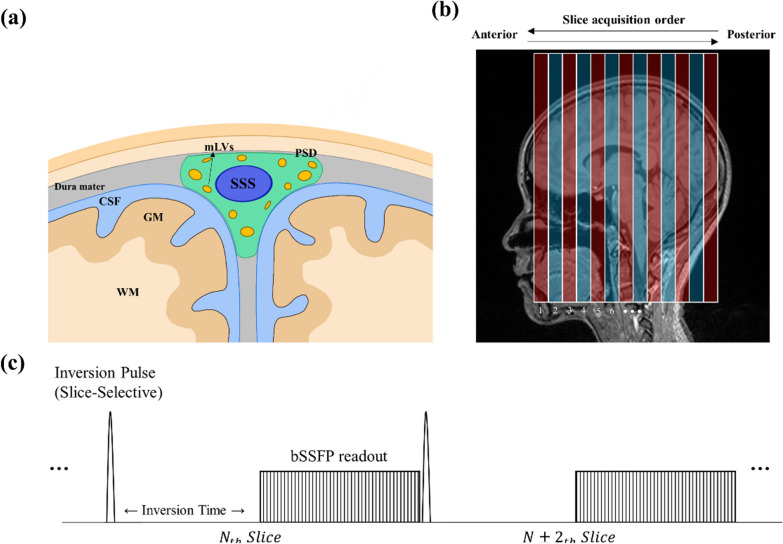
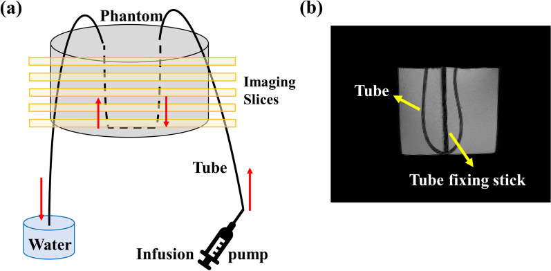
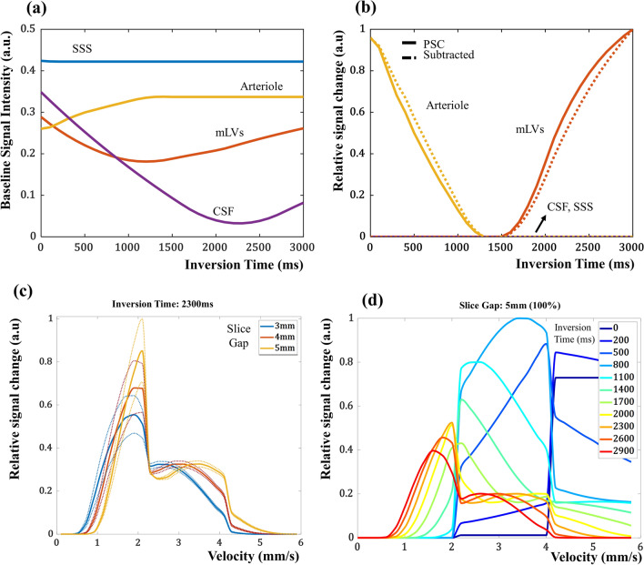
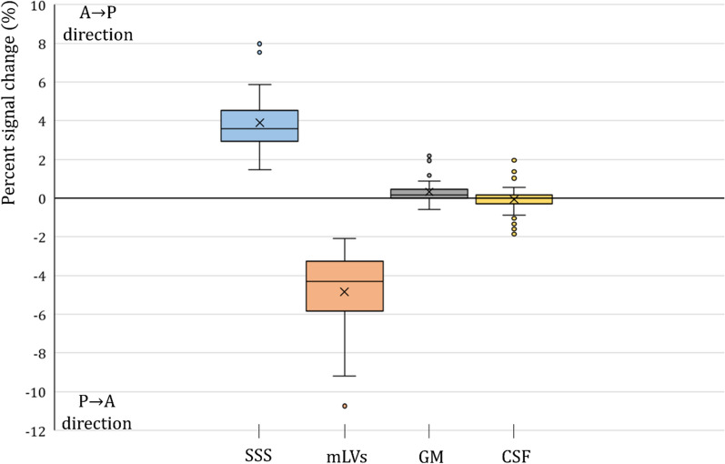
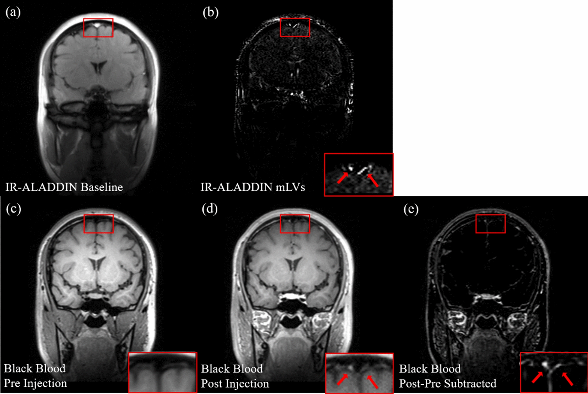
The clearance pathways of brain waste products in humans are still under debate in part due to the lack of noninvasive imaging techniques for meningeal lymphatic vessels (mLVs). In this study, we propose a new noninvasive mLVs imaging technique based on an inter-slice blood perfusion MRI called alternate ascending/descending directional navigation (ALADDIN). ALADDIN with inversion recovery (IR) at single inversion time of 2300 ms (single-TI IR-ALADDIN) clearly demonstrated parasagittal mLVs around the human superior sagittal sinus (SSS) with better detectability and specificity than the previously suggested noninvasive imaging techniques. While in many studies it has been difficult to detect mLVs and confirm their signal source noninvasively, the detection of mLVs in this study was confirmed by their posterior to anterior flow direction and their velocities and morphological features, which were consistent with those from the literature. In addition, IR-ALADDIN was compared with contrast-enhanced black blood imaging to confirm the detection of mLVs and its similarity. For the quantification of flow velocity of mLVs, IR-ALADDIN was performed at three inversion times of 2000, 2300, and 2600 ms (three-TI IR-ALADDIN) for both a flow phantom and humans. For this preliminary result, the flow velocity of the dorsal mLVs in humans ranged between 2.2 and 2.7 mm/s. Overall, (i) the single-TI IR-ALADDIN can be used as a novel non-invasive method to visualize mLVs in the whole brain with scan time of ~ 17 min and (ii) the multi-TI IR-ALADDIN can be used as a way to quantify the flow velocity of mLVs with a scan time of ~ 10 min (or shorter) in a limited coverage. Accordingly, the suggested approach can be applied to noninvasively studying meningeal lymphatic flows in general and also understanding the clearance pathways of waste production through mLVs in humans, which warrants further investigation.
Keywords: ALADDIN; Flow velocity; Magnetic resonance imaging; Meningeal lymphatic vessel; Non-invasive; Parasagittal; Perfusion imaging.
© 2023. The Author(s).
Conflict of interest statement
The authors declare no competing interests.
Figures


Similar articles
-
Feasibility of Quantifying Arterial Cerebral Blood Volume Using Multiphase Alternate Ascending/Descending Directional Navigation (ALADDIN).PLoS One. 2016 Jun 3;11(6):e0156687. doi: 10.1371/journal.pone.0156687. eCollection 2016. PLoS One. 2016. PMID: 27257674 Free PMC article.
-
Imaging of the meningeal lymphatic network in healthy adults: A 7T MRI study.J Neuroradiol. 2023 Jun;50(4):369-376. doi: 10.1016/j.neurad.2023.03.002. Epub 2023 Mar 12. J Neuroradiol. 2023. PMID: 36918053 Free PMC article.
-
Evaluation of glymphatic-meningeal lymphatic system with intravenous gadolinium-based contrast-enhancement in cerebral small-vessel disease.Eur Radiol. 2023 Sep;33(9):6096-6106. doi: 10.1007/s00330-023-09796-6. Epub 2023 Jul 6. Eur Radiol. 2023. PMID: 37410111
-
The Glymphatic System: A Review of the Challenges in Visualizing its Structure and Function with MR Imaging.Magn Reson Med Sci. 2022 Mar 1;21(1):182-194. doi: 10.2463/mrms.rev.2020-0122. Epub 2020 Nov 27. Magn Reson Med Sci. 2022. PMID: 33250472 Free PMC article. Review.
-
The Underlying Role of the Glymphatic System and Meningeal Lymphatic Vessels in Cerebral Small Vessel Disease.Biomolecules. 2022 May 25;12(6):748. doi: 10.3390/biom12060748. Biomolecules. 2022. PMID: 35740873 Free PMC article. Review.
Cited by
-
Immune cells as messengers from the CNS to the periphery: the role of the meningeal lymphatic system in immune cell migration from the CNS.Front Immunol. 2023 Aug 17;14:1233908. doi: 10.3389/fimmu.2023.1233908. eCollection 2023. Front Immunol. 2023. PMID: 37662908 Free PMC article. Review.
-
Role of meningeal lymphatic vessels in brain homeostasis.Front Immunol. 2025 Jun 19;16:1593630. doi: 10.3389/fimmu.2025.1593630. eCollection 2025. Front Immunol. 2025. PMID: 40612953 Free PMC article.
-
CSF pulsations measured in Parkinson's disease patients using EPI-based fMRI data.Front Aging Neurosci. 2024 Apr 26;16:1369522. doi: 10.3389/fnagi.2024.1369522. eCollection 2024. Front Aging Neurosci. 2024. PMID: 38737587 Free PMC article.
-
Glymphatic-lymphatic coupling: assessment of the evidence from magnetic resonance imaging of humans.Cell Mol Life Sci. 2024 Mar 13;81(1):131. doi: 10.1007/s00018-024-05141-2. Cell Mol Life Sci. 2024. PMID: 38472405 Free PMC article. Review.
-
Long-term physical exercise facilitates putative glymphatic and meningeal lymphatic vessel flow in humans.Nat Commun. 2025 Apr 9;16(1):3360. doi: 10.1038/s41467-025-58726-1. Nat Commun. 2025. PMID: 40204790 Free PMC article.
References
MeSH terms
Grants and funding
LinkOut - more resources
Full Text Sources
Medical
