

Understanding the Wnt Signaling Pathway in Acute Myeloid Leukemia Stem Cells: A Feasible Key against Relapses
- PMID: 37237497
- PMCID: PMC10215262
- DOI: 10.3390/biology12050683
Understanding the Wnt Signaling Pathway in Acute Myeloid Leukemia Stem Cells: A Feasible Key against Relapses
Abstract
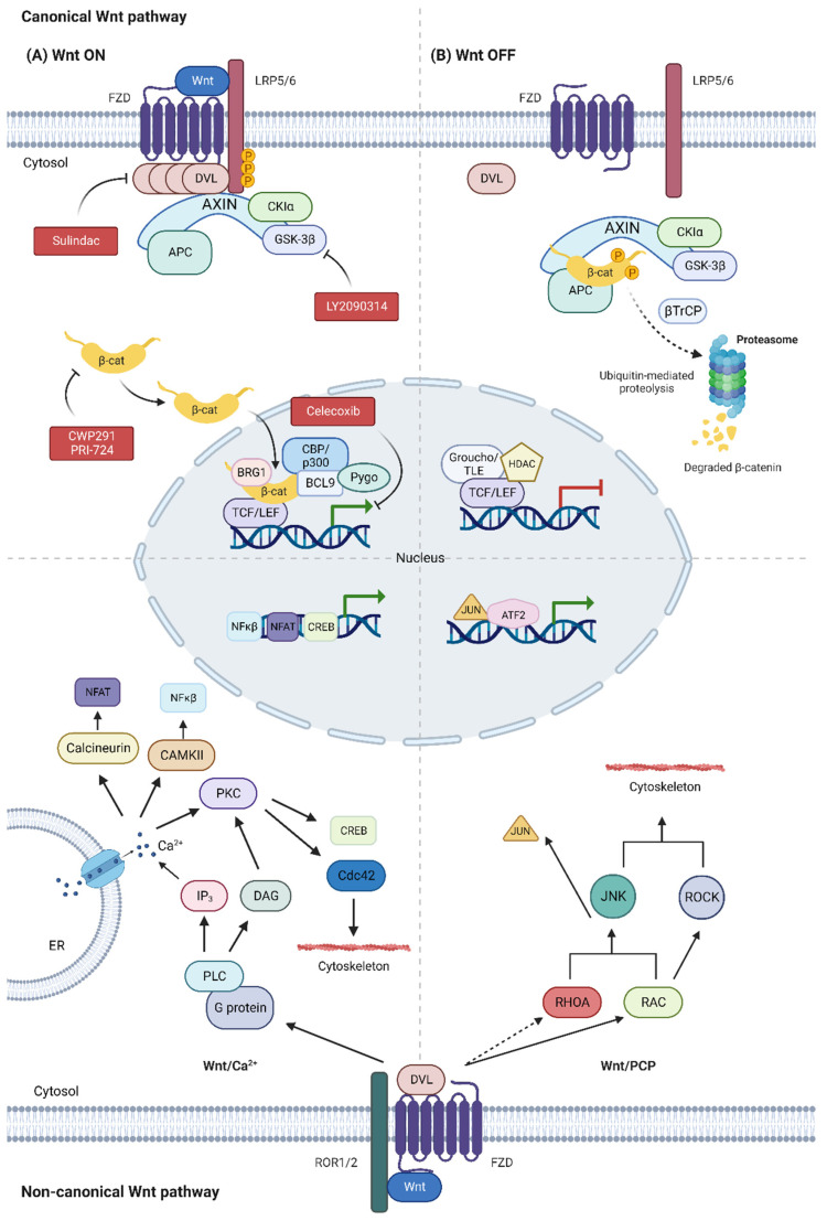
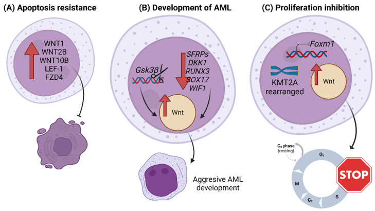
Wnt signaling is a highly conserved pathway in evolution which controls important processes such as cell proliferation, differentiation and migration, both in the embryo and in the adult. Dysregulation of this pathway can favor the development of different types of cancer, such as acute myeloid leukemia and other hematological malignancies. Overactivation of this pathway may promote the transformation of pre-leukemic stem cells into acute myeloid leukemia stem cells, as well as the maintenance of their quiescent state, which confers them with self-renewal and chemoresistance capacity, favoring relapse of the disease. Although this pathway participates in the regulation of normal hematopoiesis, its requirements seem to be greater in the leukemic stem cell population. In this review, we explore the possible therapeutic targeting of Wnt to eradicate the LSCs of AML.
Keywords: AML; HSCs; Hedgehog; LSCs; Notch; hematopoiesis; quiescence; β-catenin.
Conflict of interest statement
The authors declare no conflict of interest. Juan Manuel Alonso-Dominguez received research funding from Incyte Corporation, Pfizer International, Celgene International and Astellas Pharma Inc.
Figures
Similar articles
-
Understanding the Notch Signaling Pathway in Acute Myeloid Leukemia Stem Cells: From Hematopoiesis to Neoplasia.Cancers (Basel). 2022 Mar 12;14(6):1459. doi: 10.3390/cancers14061459. Cancers (Basel). 2022. PMID: 35326610 Free PMC article. Review.
-
[TIM-3 signaling hijacks the canonical Wnt/β-catenin pathway to maintain cancer stemness in human acute myeloid leukemia].Rinsho Ketsueki. 2023;64(6):547-552. doi: 10.11406/rinketsu.64.547. Rinsho Ketsueki. 2023. PMID: 37407480 Japanese.
-
Phytochemical Modulation of Apoptosis and Autophagy: Strategies to Overcome Chemoresistance in Leukemic Stem Cells in the Bone Marrow Microenvironment.Int Rev Neurobiol. 2017;135:249-278. doi: 10.1016/bs.irn.2017.02.012. Epub 2017 Apr 12. Int Rev Neurobiol. 2017. PMID: 28807161 Review.
-
TIM-3 signaling hijacks the canonical Wnt/β-catenin pathway to maintain cancer stemness in acute myeloid leukemia.Blood Adv. 2023 May 23;7(10):2053-2065. doi: 10.1182/bloodadvances.2022008405. Blood Adv. 2023. PMID: 36745103 Free PMC article.
-
Recreating the Bone Marrow Microenvironment to Model Leukemic Stem Cell Quiescence.Front Cell Dev Biol. 2021 Sep 13;9:662868. doi: 10.3389/fcell.2021.662868. eCollection 2021. Front Cell Dev Biol. 2021. PMID: 34589478 Free PMC article.
Cited by
-
Identifying acute myeloid leukemia subtypes based on pathway enrichment.Front Pharmacol. 2025 Mar 21;16:1557112. doi: 10.3389/fphar.2025.1557112. eCollection 2025. Front Pharmacol. 2025. PMID: 40191420 Free PMC article.
-
"Galectin-9: A double-edged sword in Acute Myeloid Leukemia".Ann Hematol. 2025 Jun;104(6):3077-3090. doi: 10.1007/s00277-025-06387-x. Ann Hematol. 2025. PMID: 40341460 Free PMC article. Review.
-
RNA modification in normal hematopoiesis and hematologic malignancies.MedComm (2020). 2024 Oct 23;5(11):e787. doi: 10.1002/mco2.787. eCollection 2024 Nov. MedComm (2020). 2024. PMID: 39445003 Free PMC article. Review.
-
Casein Kinase 2 (CK2): A Possible Therapeutic Target in Acute Myeloid Leukemia.Cancers (Basel). 2023 Jul 21;15(14):3711. doi: 10.3390/cancers15143711. Cancers (Basel). 2023. PMID: 37509370 Free PMC article. Review.
-
Therapeutic Potential of Natural Compounds to Modulate WNT/β-Catenin Signaling in Cancer: Current State of Art and Challenges.Int J Mol Sci. 2024 Nov 28;25(23):12804. doi: 10.3390/ijms252312804. Int J Mol Sci. 2024. PMID: 39684513 Free PMC article. Review.
References
-
- Khoury J.D., Solary E., Abla O., Akkari Y., Alaggio R., Apperley J.F., Bejar R., Berti E., Busque L., Chan J.K.C., et al. The 5th Edition of the World Health Organization Classification of Haematolymphoid Tumours: Myeloid and Histiocytic/Dendritic Neoplasms. Leukemia. 2022;36:1703–1719. doi: 10.1038/s41375-022-01613-1. - DOI - PMC - PubMed
-
- Arber D.A., Orazi A., Hasserjian R.P., Borowitz M.J., Calvo K.R., Kvasnicka H.-M., Wang S.A., Bagg A., Barbui T., Branford S., et al. International Consensus Classification of Myeloid Neoplasms and Acute Leukemias: Integrating Morphologic, Clinical, and Genomic Data. Blood. 2022;140:1200–1228. doi: 10.1182/blood.2022015850. - DOI - PMC - PubMed
Publication types
LinkOut - more resources
Full Text Sources
