. 2023 May 16;12(5):725.
doi: 10.3390/biology12050725.

Contrariety of Human Bone Marrow Mesenchymal Stromal Cell Functionality in Modulating Circulatory Myeloid and Plasmacytoid Dendritic Cell Subsets

Affiliations

Contrariety of Human Bone Marrow Mesenchymal Stromal Cell Functionality in Modulating Circulatory Myeloid and Plasmacytoid Dendritic Cell Subsets

Crystal C Uwazie et al. Biology (Basel). .

Abstract

Mesenchymal Stromal Cells (MSCs) derived from bone marrow are widely tested in clinical trials as a cellular therapy for potential inflammatory disorders. The mechanism of action of MSCs in mediating immune modulation is of wide interest. In the present study, we investigated the effect of human bone-marrow-derived MSCs in modulating the circulating peripheral blood dendritic cell responses through flow cytometry and multiplex secretome technology upon their coculture ex vivo. Our results demonstrated that MSCs do not significantly modulate the responses of plasmacytoid dendritic cells. However, MSCs dose-dependently promote the maturation of myeloid dendritic cells. Mechanistic analysis showed that dendritic cell licensing cues (Lipopolysaccharide and Interferon-gamma) stimulate MSCs to secret an array of dendritic cell maturation-associated secretory factors. We also identified that MSC-mediated upregulation of myeloid dendritic cell maturation is associated with the unique predictive secretome signature. Overall, the present study demonstrated the dichotomy of MSC functionality in modulating myeloid and plasmacytoid dendritic cells. This study provides clues that clinical trials need to investigate if circulating dendritic cell subsets in MSC therapy can serve as potency biomarkers.

Keywords: circulating dendritic cells; immunomodulation; mesenchymal stromal cells; secretome.


Conflict of interest statement

The authors declare that they have no competing interest.

Figures

Figure 1
Figure 1
Characteristics of circulating dendritic cells with and without LPS and IFNγ stimulation. Dendritic-cell-enriched PBMCs were stimulated with LPS and IFNγ for 24 h. Subsequently, dendritic cell maturation was measured by flow cytometry and secretory analytes were quantified in the supernatants using Luminex xMAP (multi-analyte profiling) technology. (A) Representative flow cytometry plot and gating strategy are shown to identify plasmacytoid (Lineage−(CD3−CD14−CD16−CD56−CD19−CD20−) HLADR+CD303+CD1C−) and myeloid (Lineage−(CD3−CD14−CD16−CD56−CD19−CD20−) HLADR+CD303−CD1C+) dendritic cell subtypes. CD83 expression between−and + stim conditions are shown for (B,C) Lineage−HLADR+CD303+CD1C− and (D,E) Lineage−HLADR+CD303−CD1C+ populations with representative and cumulative plots. (F) Twnety-nine secretory analytes are organized based on their magnitude of differences between stim and no stim conditions. The magnitude of secretory difference is calculated as the geometric mean of the ratio between stim and no stim condition with 95% confidence interval. (G) concentrations of individual analytes are shown between stim (+) and no stim (−) cultures with mean and standard deviation. Statistical significance is shown as ns, p > 0.05; * p ≤ 0.05; ** p ≤ 0.01; *** p ≤ 0.001; **** p ≤ 0.0001. M indicates media control. Cumulative results are shown from seven independent donors.
Figure 2
Figure 2
Immunophenotype of human bone-marrow-derived mesenchymal stromal cells. The identity of human bone-marrow-derived MSCs is confirmed in flow cytometry. Histogram plots are shown with appropriate isotype controls (blue) and marker (red) expressions. The results are shown from six independent donors.
Figure 3
Figure 3
Dose-dependent effect of LPS and IFNγ stimulation of human bone marrow MSCs. MSCs derived from five independent donors were seeded at the identical cell density (10,000 cells/well in 96-well plate) and stimulated with the indicated concentrations of LPS and IFNγ. Forty-eight hours later, supernatants were centrifuged and stored. Secretory analytes were quantified in the supernatants using Luminex xMAP (multi-analyte profiling) technology. Twenty-nine analytes are hierarchically organized based on their magnitude of response to the stimulus LPS and IFNγ. The magnitude of upregulation and 95% confidence interval is shown for each analyte. Statistical significance is shown as ns, p > 0.05; * p ≤ 0.05; ** p ≤ 0.01; *** p ≤ 0.001; **** p ≤ 0.0001. M indicates media control. Cumulative results are shown from five independent donors.
Figure 4
Figure 4
Effect of human bone marrow MSCs on the maturation of dendritic cell subsets. Human bone-marrow-derived MSCs were seeded at different cell densities. Subsequently, dendritic-cell-enriched PBMCs, stimulated with LPS (100 ng/mL) and IFNγ (40 ng/mL) were cocultured with MSCs. Twenty-four hours post incubation, they were stained with antibodies for lineage, HLADR, CD303, CD1C and CD83 and acquired in flow cytometer. Representative flow cytometry plots of (A) Lineage-(CD3−CD14−CD16−CD56−CD19−CD20−) HLADR+CD303−CD1C+ and (B) Lineage− (CD3−CD14−CD16−CD56−CD19−CD20−) HLADR+CD303+CD1C- are shown for the expression of CD83 with and without MSCs. Spaghetti plots depict the frequencies of (C) Lineage−HLADR+CD303−CD1C+CD83+ and (D) Lineage−HLADR+CD303+CD1C−CD83+ populations in the presence of various doses of MSCs. Dose dependence values, statistical significance and 95% confidence interval are shown in individual plots. Cumulative data derived from six independent MSC and seven PBMC donors are shown.
Figure 5
Figure 5
Dose-dependent effect of MSCs in modulating the secretome of LPS- and IFNγ-activated DC enriched PBMCs. Supernatants derived from the cocultures of LPS and IFNγ stimulated dendritic-cell-enriched PBMCs and MSCs were analyzed for 30-plex secretome using Luminex xMAP (multi-analyte profiling) technology. Spaghetti plots depict the levels of individual secretory molecules (pg/mL) in MSC dose-dependent cocultures with dendritic-cell-enriched PBMCs. Statistically significant secretory molecules that are upregulated with the increasing doses of MSCs in the cocultures are highlighted in blue. In contrast, statistically significant secretory molecules that are downregulated with the increasing doses of MSCs in the cocultures are highlighted in orange. Statistically non-significant molecules are depicted in black. M stands for Media only control (No MSCs and No DC-enriched PBMCs). Dose dependency values and statistical significance are shown in each graph (ns, p > 0.05; * p ≤ 0.05; ** p ≤ 0.01; *** p ≤ 0.001; **** p ≤ 0.0001). IFNγ in the 30-plex is excluded in the analysis since it is present in the inoculum. Cumulative data derived from six independent MSC and seven PBMC donors are shown.
Figure 6
Figure 6
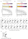
Correlation patterns between MSC-induced frequency of dendritic cell subsets and secretome. Quantitative levels of individual secretory molecules derived from the cocultures of MSCs and dendritic-cell-enriched PBMCs were subjected to linear regression analysis with the corresponding percentage of (A) Lineage-HLADR+CD303-CD1C+CD83+ and (B) Lineage-HLADR+CD303+CD1C-CD83+ populations. Correlation coefficient r-values of 1 and 0 specify the best direct and no correlations, respectively. The statistical significance of each of these correlations is shown with the heat map that describes the negative logarithmic of p-values. Concentration secretory analytes were transformed to logarithmic values to fit linear regression curves. Statistically high significant correlations with (C) Lineage-HLADR+CD303-CD1C+CD83+ and (D) Lineage-HLADR+CD303+CD1C-CD83+ populations are shown in the individual plots. Cumulative data derived from six independent MSC and seven PBMC donors are used in the linear regression analysis.
Figure 7
Figure 7
Effect of human bone-marrow-derived MSCs in modulating IFNα production in plasmacytoid dendritic cells. PBMCs (100,000 cells/well) were cocultured in the presence and absence of MSCs (10,000 cells/well) and were stimulated with R837 (20 μg/mL). The cells were incubated with BD Golgi Plug for 14 h and subsequently stained with surface antibodies for Lineage, HLADR, CD303, CD1C and intracellular IFNα. (A) Representative flow plots describing the IFNα production in Lineage-HLADR+CD303-CD1C+ and Lineage-HLADR+CD303+CD1C- populations are shown. (B) The effect of MSCs in the frequency of Lineage-HLADR+CD303+CD1C- IFNα+ populations are shown with a representative flow plot and (C) cumulative graphs. (C) Cumulative data derived from five independent MSC and five PBMC donors are shown. Statistical significance is shown. ns = Nonsignificant.

References

    1. Galipeau J., Sensebe L. Mesenchymal Stromal Cells: Clinical Challenges and Therapeutic Opportunities. Cell Stem Cell. 2018;22:824–833. doi: 10.1016/j.stem.2018.05.004. - DOI - PMC - PubMed
    1. Pittenger M.F., Discher D.E., Peault B.M., Phinney D.G., Hare J.M., Caplan A.I. Mesenchymal stem cell perspective: Cell biology to clinical progress. NPJ Regen. Med. 2019;4:22. doi: 10.1038/s41536-019-0083-6. - DOI - PMC - PubMed
    1. Moll G., Ankrum J.A., Kamhieh-Milz J., Bieback K., Ringden O., Volk H.D., Geissler S., Reinke P. Intravascular Mesenchymal Stromal/Stem Cell Therapy Product Diversification: Time for New Clinical Guidelines. Trends Mol. Med. 2019;25:149–163. doi: 10.1016/j.molmed.2018.12.006. - DOI - PubMed
    1. Kabat M., Bobkov I., Kumar S., Grumet M. Trends in mesenchymal stem cell clinical trials 2004–2018: Is efficacy optimal in a narrow dose range? Stem Cells Transl. Med. 2020;9:17–27. doi: 10.1002/sctm.19-0202. - DOI - PMC - PubMed
    1. Moll G., Hoogduijn M.J., Ankrum J.A. Editorial: Safety, Efficacy and Mechanisms of Action of Mesenchymal Stem Cell Therapies. Front. Immunol. 2020;11:243. doi: 10.3389/fimmu.2020.00243. - DOI - PMC - PubMed

LinkOut - more resources