. 2023 Apr 27;10(5):539.
doi: 10.3390/bioengineering10050539.

Finite Element Modeling of Residual Hearing after Cochlear Implant Surgery in Chinchillas

Affiliations

Finite Element Modeling of Residual Hearing after Cochlear Implant Surgery in Chinchillas

Nicholas Castle et al. Bioengineering (Basel). .

Abstract

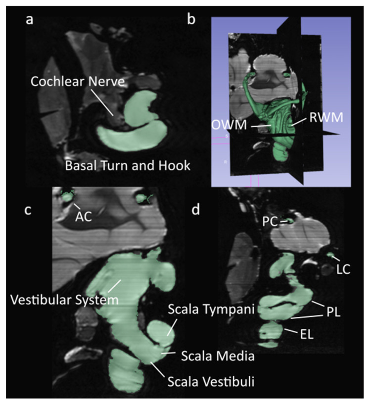
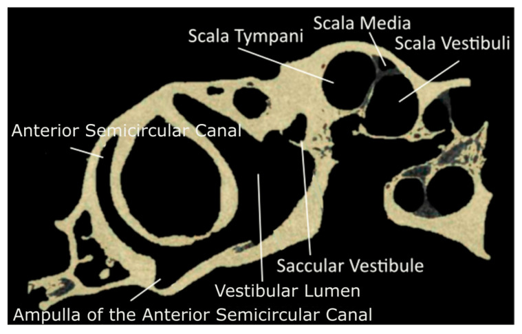
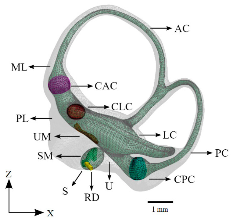
Cochlear implant (CI) surgery is one of the most utilized treatments for severe hearing loss. However, the effects of a successful scala tympani insertion on the mechanics of hearing are not yet fully understood. This paper presents a finite element (FE) model of the chinchilla inner ear for studying the interrelationship between the mechanical function and the insertion angle of a CI electrode. This FE model includes a three-chambered cochlea and full vestibular system, accomplished using µ-MRI and µ-CT scanning technologies. This model's first application found minimal loss of residual hearing due to insertion angle after CI surgery, and this indicates that it is a reliable and helpful tool for future applications in CI design, surgical planning, and stimuli setup.

Keywords: chinchilla; cochlear implant; finite element; inner ear; insertion angle.


Conflict of interest statement

The authors declare no conflict of interest. The terms of this arrangement are being managed in accordance with University of Oklahoma policies on conflict of interest.

Figures

Figure 1
Figure 1
Segmented µMRI scans of the chinchilla subject with key structures labeled. The lymphatic fluid of the inner ear is shown in green. (a) Transverse plane; (b) 3D view of the entire segmentation; (c) Saggital plane; (d) Coronal plane. Refer to Table 1 for symbol definitions.
Figure 2
Figure 2
Segmented µCT scan in the sagittal plane of the chinchilla subject with key structures labeled. Refer to Table 1 for symbol definitions.
Figure 3
Figure 3
The coordinate system used in all imaging for this paper. The x and z axes are held in the sagittal plane as if viewed from the subject’s left side.
Figure 4
Figure 4
The vestibular system of the computational model. The saccule, utricle, and semicircular canals appear as a continuous volume of lymphatic fluid (green). The sensory organs of the vestibular system are also shown. Refer to Table 1 for symbol definitions.
Figure 5
Figure 5
The cochlea of the computational model without the cochlear implant. The design of the basilar membrane (red) is apparent as a ribbon with varying width and thickness, attached on both sides to bony supports (grey). The distal side of the basilar membrane is visible as the attachment point for the end of the Reissner’s membrane. Refer to Table 1 for symbol definitions.
Figure 6
Figure 6
The full meshed model of the cochlea is presented with the length of the cochlear implant electrode inserted. (a) Cross-sections of the base (1) and apex (2) ends of the cochlear implant electrode; (b) The path of the cochlear implant electrode through the scala tympani of the meshed model.
Figure 7
Figure 7
Displacement of the basilar membrane from the base to the apex of the cochlea before insertion of the CI (400 Hz: black, 1000 Hz: orange, 2000 Hz: yellow, 4000 Hz: green, 6000 Hz: blue, 8000 Hz: purple, 10,000 Hz: grey). Model input was the experimentally determined frequency dependent displacement of the stapes at 90 dB [42]. (a,b) show unnormalized displacement of the basilar membrane and basilar membrane displacement normalized with that of the stapes footplate, respectively. (c) shows the location of the maximum displacement for the model (black) at each frequency compared to an experimentally obtained benchmark [54].
Figure 8
Figure 8
Displacement of the basilar membrane from the base to the apex of the cochlea at each evaluated insertion angle of the cochlear electrode (400 Hz: black, 1000 Hz: orange, 2000 Hz: yellow, 4000 Hz: green, 6000 Hz: blue, 8000 Hz: purple, 10,000 Hz: grey). Model input was the experimentally determined frequency dependent displacement of the stapes at 90 dB [42]. The upper figure in each set and the lower set show unnormalized displacement of the basilar membrane and basilar membrane displacement normalized with that of the stapes footplate, respectively.

Similar articles

Cited by

References

    1. World Health Organization Deafness and Hearing Loss. [(accessed on 1 January 2022)]. Available online: https://www.who.int/news-room/fact-sheets/detail/deafness-and-hearing-loss.
    1. Olze H., Szczepek A.J., Haupt H., Förster U., Zirke N., Gräbel S., Mazurek B. Cochlear Implantation Has a Positive Influence on Quality of Life, Tinnitus, and Psychological Comorbidity. Laryngoscope. 2011;121:2220–2227. doi: 10.1002/lary.22145. - DOI - PubMed
    1. Sousa A.F.D., Couto M.I.V., Martinho-Carvalho A.C. Quality of Life and Cochlear Implant: Results in Adults with Postlingual Hearing Loss. Braz. J. Otorhinolaryngol. 2018;84:494–499. doi: 10.1016/j.bjorl.2017.06.005. - DOI - PMC - PubMed
    1. Hofmann M., Meloche M., Zwolan T.A. Health Related Quality of Life in Adolescent Cochlear Implant Users. Cochlear Implant. Int. 2020;21:198–205. doi: 10.1080/14670100.2020.1724676. - DOI - PubMed
    1. Issing C., Baumann U., Pantel J., Stöver T. Cochlear Implant Therapy Improves the Quality of Life in Older Patients—A Prospective Evaluation Study. Otol. Neurotol. 2020;41:1214–1221. doi: 10.1097/MAO.0000000000002741. - DOI - PubMed