Review
. 2023 May 18;10(5):606.
doi: 10.3390/bioengineering10050606.

3D Bioprinting for Vascularization

Affiliations
Review

3D Bioprinting for Vascularization

Amatullah Mir et al. Bioengineering (Basel). .

Abstract

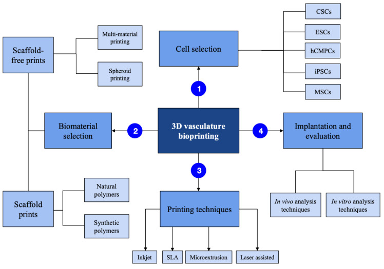
In the world of clinic treatments, 3D-printed tissue constructs have emerged as a less invasive treatment method for various ailments. Printing processes, scaffold and scaffold free materials, cells used, and imaging for analysis are all factors that must be observed in order to develop successful 3D tissue constructs for clinical applications. However, current research in 3D bioprinting model development lacks diverse methods of successful vascularization as a result of issues with scaling, size, and variations in printing method. This study analyzes the methods of printing, bioinks used, and analysis techniques in 3D bioprinting for vascularization. These methods are discussed and evaluated to determine the most optimal strategies of 3D bioprinting for successful vascularization. Integrating stem and endothelial cells in prints, selecting the type of bioink according to its physical properties, and choosing a printing method according to physical properties of the desired printed tissue are steps that will aid in the successful development of a bioprinted tissue and its vascularization.

Keywords: 3D modeling; 3D printing; 3D tissues; 3D-printed sensors; bioinks; biomanufacturing; biomechanics; bioprinting; bone; hard tissue; oral diseases; organ-on-a-chip; scaffolds; skin; soft tissue; spheroids; stem cells; tissue engineering; vascularization; vasculature.


Conflict of interest statement

The authors declare no conflict of interest.

Figures

Figure 1
Figure 1
3-dimensional bioprinting process summarized.
Figure 2
Figure 2
There are four methods of bioprinting: inkjet (A), microextrusion (B), laser-assisted (C), and stereolithographic (D).
Figure 3
Figure 3
Schematic of the laser-assisted bioprinting (LAB) fabrication method and its direct printing onto a receiving substrate. Adapted from Keriquel et al. [8].
Figure 4
Figure 4
3D Imaris software analysis of 3D bioprinted patch stained with antibodies against CD31 (green) and Hoechst stain (blue). (A,B) 3D rendering of the endothelial structure of within the 3D bioprinted patch. Adapted from Gentile et al. [52].
Figure 5
Figure 5
Natural and synthetic polymer properties, summarized.
Figure 6
Figure 6
Spheroid aspiration, transfer, and deposition into a support hydrogel. Adapted from Daly et al. [60].
Figure 7
Figure 7
Pluronic, thrombin, and cell-laden inks, which contain gelatin, fibrinogen, and cells, are printed in a 3D perfusion chip, which is cast with ECM material. Thrombin and fibrinogen polymerize to form fibrin, and the system is perfused with an external pump. Adapted from Kolesky et al. [13].

References

    1. Datta P., Ayan B., Ozbolat I.T. Bioprinting for vascular and vascularized tissue biofabrication. Acta Biomater. 2017;51:1–20. doi: 10.1016/j.actbio.2017.01.035. - DOI - PubMed
    1. Laschke M.W., Menger M.D. Spheroids as vascularization units: From angiogenesis research to tissue engineering applications. Biotechnol. Adv. 2017;35:782–791. doi: 10.1016/j.biotechadv.2017.07.002. - DOI - PubMed
    1. Cui H., Miao S., Esworthy T., Zhou X., Lee S.J., Liu C., Yu Z.X., Fisher J.P., Mohiuddin M., Zhang L.G. 3D bioprinting for cardiovascular regeneration and pharmacology. Adv. Drug Deliv. Rev. 2018;132:252–269. doi: 10.1016/j.addr.2018.07.014. - DOI - PMC - PubMed
    1. Lovett M., Lee K., Edwards A., Kaplan D.L. Vascularization Strategies for Tissue Engineering. Tissue Eng. Part B Rev. 2009;15:353–370. doi: 10.1089/ten.teb.2009.0085. - DOI - PMC - PubMed
    1. Tengood J.E., Ridenour R., Brodsky R., Russell A.J., Little S.R. Sequential Delivery of Basic Fibroblast Growth Factor and Platelet-Derived Growth Factor for Angiogenesis. Tissue Eng. Part A. 2011;17:1181–1189. doi: 10.1089/ten.tea.2010.0551. - DOI - PMC - PubMed

LinkOut - more resources