. 2023 May 11;12(5):1082.
doi: 10.3390/antiox12051082.

Aucubin Exerts Neuroprotection against Forebrain Ischemia and Reperfusion Injury in Gerbils through Antioxidative and Neurotrophic Effects

Affiliations

Aucubin Exerts Neuroprotection against Forebrain Ischemia and Reperfusion Injury in Gerbils through Antioxidative and Neurotrophic Effects

Joon Ha Park et al. Antioxidants (Basel). .

Abstract

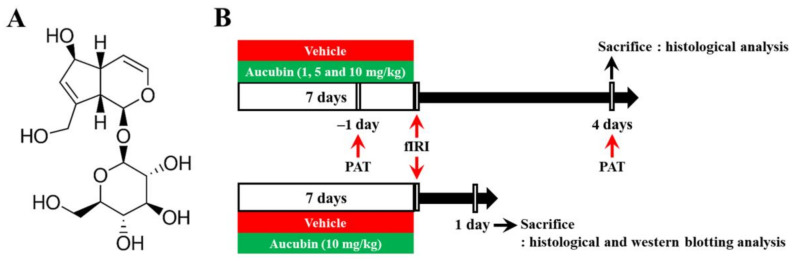
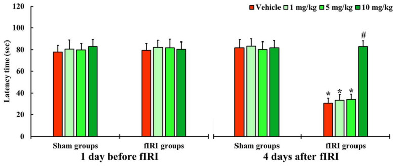
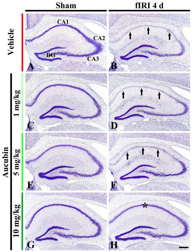
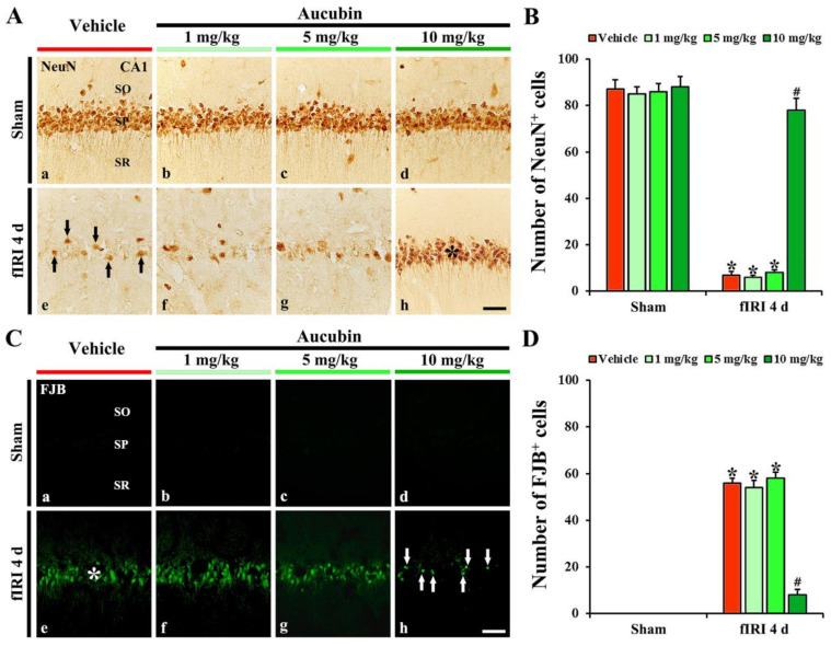
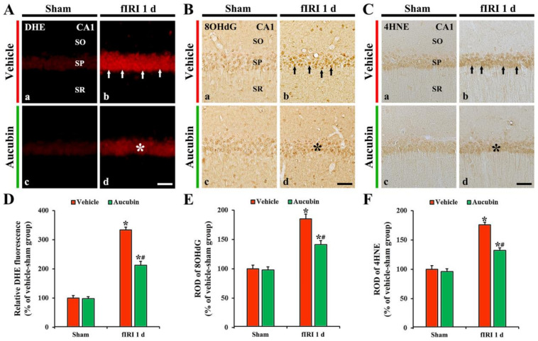
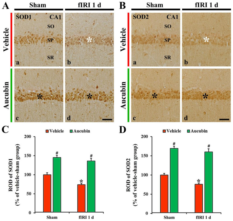
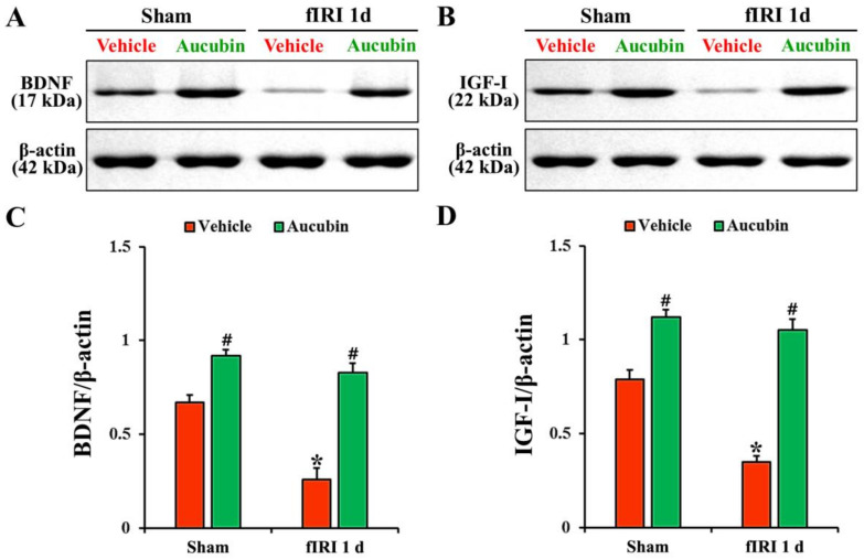
Aucubin is an iridoid glycoside that displays various pharmacological actions including antioxidant activity. However, there are few reports available on the neuroprotective effects of aucubin against ischemic brain injury. Thus, the aim of this study was to investigate whether aucubin protected against damage to hippocampal function induced by forebrain ischemia-reperfusion injury (fIRI) in gerbils, and to examine whether aucubin produced neuroprotection in the hippocampus against fIRI and to explore its mechanisms by histopathology, immunohistochemistry, and Western analysis. Gerbils were given intraperitoneal injections of aucubin at doses of 1, 5, and 10 mg/kg, respectively, once a day for seven days before fIRI. As assessed by the passive avoidance test, short-term memory function following fIRI significantly declined, whereas the decline in short-term memory function due to fIRI was ameliorated by pretreatment with 10 mg/kg, but not 1 or 5 mg/kg, of aucubin. Most of the pyramidal cells (principal cells) of the hippocampus died in the Cornu Ammonis 1 (CA1) area four days after fIRI. Treatment with 10 mg/kg, but not 1 or 5 mg/kg, of aucubin protected the pyramidal cells from IRI. The treatment with 10 mg/kg of aucubin significantly reduced IRI-induced superoxide anion production, oxidative DNA damage, and lipid peroxidation in the CA1 pyramidal cells. In addition, the aucubin treatment significantly increased the expressions of superoxide dismutases (SOD1 and SOD2) in the pyramidal cells before and after fIRI. Furthermore, the aucubin treatment significantly enhanced the protein expression levels of neurotrophic factors, such as brain-derived neurotrophic factor and insulin-like growth factor-I, in the hippocampal CA1 area before and after IRI. Collectively, in this experiment, pretreatment with aucubin protected CA1 pyramidal cells from forebrain IRI by attenuating oxidative stress and increasing neurotrophic factors. Thus, pretreatment with aucubin can be a promising candidate for preventing brain IRI.

Keywords: aucubin; hippocampus; histopathology; immunohistochemistry; oxidative stress; transient ischemia; western blotting.


Conflict of interest statement

The authors declare that they have no conflict of interest.

Figures

Figure 1
Figure 1
(A) The chemical structure of aucubin and (B) schematic representation of the experimental procedure. Aucubin was dissolved in saline and intraperitoneally injected once/day for 7 days before fIRI. Passive avoidance test (PAT) was conducted 1 day before and 4 days after fIRI. The gerbils were sacrificed to examine neuroprotection on day 1 and to investigate the mechanisms of the neuroprotection 4 days after fIRI.
Figure 2
Figure 2
Latency time by PAT in the vehicle-sham, vehicle-fIRI, 1 mg/kg aucubin-sham, 1 mg/kg aucubin-fIRI, 5 mg/kg aucubin-sham, 5 mg/kg aucubin-fIRI, 10 mg/kg aucubin-sham, and 10 mg/kg aucubin-fIRI groups 1 day before and 4 days after fIRI. In the vehicle-fIRI, 1 and 5 mg/kg aucubin-fIRI groups, the latency times are significantly shortened when compared with the vehicle-sham group. On the other hand, in the 10 mg/kg aucubin-fIRI group, the latency time is similar to that evaluated in the sham-vehicle group. Values are expressed in mean ± SEM (n = 7, respectively; * p < 0.05 vs. each sham group, # p < 0.05 vs. vehicle-fIRI group).
Figure 3
Figure 3
Representative images of Nissl staining in the hippocampus of the vehicle-sham (A), vehicle-fIRI (B), 1 mg/kg aucubin-sham (C), 1 mg/kg aucubin-fIRI (D), 5 mg/kg aucubin-sham (E), 5 mg/kg aucubin-fIRI (F), 10 mg/kg aucubin-sham (G), and 10 mg/kg aucubin-fIRI (H) groups at 4 days after fIRI. Note that CV stainability in the stratum pyramidale (arrows in (B,D,F)) of the CA1 area in the vehicle-fIRI, 1 mg/kg aucubin-fIRI, and 5 mg/kg aucubin-fIRI groups is pale after fIRI. However, in the 10 mg/kg aucubin fIRI group, strong CV stainability is shown in the CA1 stratum pyramidale (asterisk in (H)). CA, cornu ammonis; DG, dentate gyrus. Scale bar = 200 μm.
Figure 4
Figure 4
(A,C) Representative images of NeuN immunohistochemistry (A) and FJB fluorescence staining (C) in the CA1 area of the vehicle-sham (A(a),C(a)), vehicle-fIRI (A(e),C(e)), 1 mg/kg aucubin-sham (A(b),C(b)), 1 mg/kg aucubin-fIRI (A(f),C(f)), 5 mg/kg aucubin-sham (A(c),C(c)), 5 mg/kg aucubin-fIRI (A(g),C(g)), 10 mg/kg aucubin-sham (A(d),C(d)), and 10 mg/kg aucubin-fIRI (A(h),C(h)) groups at 4 days after fIRI. In the vehicle-fIRI group, a few NeuN+ (black arrows in (A(e))) and many FJB+ (white asterisk in (C(e))) pyramidal neurons in the stratum pyramidale (SP) are shown, whereas, in the 10 mg/kg aucubin-fIRI group, abundant NeuN+ (black asterisk in (A(h))) and a few FJB+ (white arrows in (C(h))) pyramidal neurons are found in the SP. SO, stratum oriens; SR, stratum radiatum. Scale bar = 60 µm. (B,D) The mean numbers of NeuN+ (B) and FJB+ (D) pyramidal neurons in the CA1 area. Values are expressed in mean ± SEM (n = 7, respectively; * p < 0.05 vs. each sham group, # p < 0.05 vs. vehicle-fIRI group).
Figure 5
Figure 5
(AC) Representative images of DHE fluorescence staining (A), 8OHdG (B), and 4HNE (C) immunohistochemistry in the CA1 area of the vehicle-sham (A(a),B(a),C(a)), vehicle-fIRI (A(b),B(b),C(b)), aucubin-sham (A(c),B(c),C(c)), and aucubin-fIRI (A(d),B(d),C(d)) groups 1 day after fIRI. In the vehicle-fIRI group, DHE fluorescence, 8OHdG, and 4HNE immunoreactivity in CA1 pyramidal neurons (arrows in (A(b),B(b),C(b))) are dramatically increased. However, in the aucubin-fIRI group, DHE fluorescence, 8OHdG, and 4HNE immunoreactivity are significantly lower (asterisks in (A(d),B(d),C(d))) than that shown in the vehicle-fIRI group. SO, stratum oriens; SP, stratum pyramidale; SR, stratum radiatum. Scale bar = 60 µm. (DF) Quantitative analyses of DHE fluorescence intensity (D), 8OHdG (E), and 4HNE (F) in CA1 pyramidal neurons. Values are expressed in mean ± SEM (n = 7, respectively; * p < 0.05 vs. each sham group, # p < 0.05 vs. vehicle-fIRI group).
Figure 6
Figure 6
(A,B) Representative images of SOD1 (A) and SOD2 (B) immunohistochemistry in the CA1 area of the vehicle-sham (A(a),B(a)), vehicle-fIRI (A(b),B(b)), aucubin-sham (A(c),B(c)), and aucubin-fIRI (A(d),B(d)) groups 1 day after fIRI. In the vehicle-fIRI group, SOD1 and SOD2 immunoreactivity in pyramidal cells (white asterisks in (A(b),B(b)) are significantly reduced when compared with those found in the vehicle-sham group. In the aucubin-sham and aucubin-fIRI groups, SOD1 and SOD2 immunoreactivity in the pyramidal neurons (black asterisks in (A(c,d),B(c,d))) are apparently higher than those shown in the vehicle-sham groups. SO, stratum oriens; SP, stratum pyramidale; SR, stratum radiatum. Scale bar = 60 µm. (C,D) Quantitative analyses of SOD1 (C) and SOD2 (D) immunoreactivity in CA1 pyramidal neurons. Values are expressed in mean ± SEM (n = 7, respectively; * p < 0.05 vs. each sham group, # p < 0.05 vs. vehicle-fIRI group).
Figure 7
Figure 7
(A,B) Representative Western blot images of BDNF and IGF-I in the CA1 area of the vehicle-sham, vehicle-fIRI, aucubin-sham, and aucubin-fIRI groups 1 day after fIRI. In the vehicle-fIRI group, BDNF and IGF-I levels are significantly reduced when compared with those found in the vehicle-sham groups. In the aucubin-sham and aucubin-fIRI groups, BDNF and IGF-I levels are significantly higher than those observed in the vehicle-sham groups. (C,D) Quantitative analyses of BDNF and IGF-I levels by normalization to β-actin level, respectively. Values are expressed in mean ± SEM (n = 5, respectively; * p < 0.05 vs. each sham group, # p < 0.05 vs. vehicle-fIRI group).

References

    1. Frisch S., Thiel F., Schroeter M.L., Jentzsch R.T. Apathy and cognitive deficits in patients with transient global ischemia after cardiac arrest. Cogn. Behav. Neurol. Off. J. Soc. Behav. Cogn. Neurol. 2017;30:172–175. doi: 10.1097/WNN.0000000000000139. - DOI - PubMed
    1. Wen H., Liu L., Zhan L., Liang D., Li L., Liu D., Sun W., Xu E. Neuroglobin mediates neuroprotection of hypoxic postconditioning against transient global cerebral ischemia in rats through preserving the activity of na(+)/k(+) atpases. Cell Death Dis. 2018;9:635. doi: 10.1038/s41419-018-0656-0. - DOI - PMC - PubMed
    1. Lin C.S., Polsky K., Nadler J.V., Crain B.J. Selective neocortical and thalamic cell death in the gerbil after transient ischemia. Neuroscience. 1990;35:289–299. doi: 10.1016/0306-4522(90)90083-G. - DOI - PubMed
    1. Wahul A.B., Joshi P.C., Kumar A., Chakravarty S. Transient global cerebral ischemia differentially affects cortex, striatum and hippocampus in bilateral common carotid arterial occlusion (bccao) mouse model. J. Chem. Neuroanat. 2018;92:1–15. doi: 10.1016/j.jchemneu.2018.04.006. - DOI - PubMed
    1. Globus M.Y., Busto R., Martinez E., Valdes I., Dietrich W.D., Ginsberg M.D. Comparative effect of transient global ischemia on extracellular levels of glutamate, glycine, and gamma-aminobutyric acid in vulnerable and nonvulnerable brain regions in the rat. J. Neurochem. 1991;57:470–478. doi: 10.1111/j.1471-4159.1991.tb03775.x. - DOI - PubMed