. 2023 May 20;12(5):1134.
doi: 10.3390/antiox12051134.

JAC4 Alleviates Rotenone-Induced Parkinson's Disease through the Inactivation of the NLRP3 Signal Pathway

Affiliations

JAC4 Alleviates Rotenone-Induced Parkinson's Disease through the Inactivation of the NLRP3 Signal Pathway

Lu Zou et al. Antioxidants (Basel). .

Abstract

Parkinson's disease (PD) is the fastest-growing neurodegeneration disease, characterized typically by a progressive loss of dopaminergic neurons in the substantia nigra, and there are no effective therapeutic agents to cure PD. Rotenone (Rot) is a common and widely used pesticide which can directly inhibit mitochondrial complex I, leading to a loss of dopaminergic neurons. Our previous studies proved that the JWA gene (arl6ip5) may play a prominent role in resisting aging, oxidative stress and inflammation, and JWA knockout in astrocytes increases the susceptibility of mice to 1-Methyl-4-phenyl-1,2,3,6-tetrahydropyridine (MPTP)-induced PD. JWA-activating compound 4 (JAC4) is a small-molecule activator of the JWA gene, but its role in and mechanism against PD have not yet been clarified. In the present study, we showed that the JWA expression level is strongly related to tyrosine hydroxylase (TH) in different growth periods of mice. Additionally, we constructed models with Rot in vivo and in vitro to observe the neuroprotective effects of JAC4. Our results demonstrated that JAC4 prophylactic intervention improved motor dysfunction and dopaminergic neuron loss in mice. Mechanistically, JAC4 reduced oxidative stress damage by reversing mitochondrial complex I damage, reducing nuclear factor kappa-B (NF-κB) translocation and repressing nucleotide-binding domain, leucine-rich-containing family and pyrin domain-containing-3 (NLRP3) inflammasome activation. Overall, our results provide proof that JAC4 could serve as a novel effective agent for PD prevention.

Keywords: JAC4; NLRP3 inflammasome; Parkinson’s disease; mitochondrial complex I; oxidative stress; rotenone.


Conflict of interest statement

The authors declare no conflict of interest.

Figures

Figure 1
Figure 1
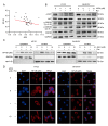
The expressions of JWA and TH are positively correlated in human and mice. (A) JWA mRNA expression was measured in a case–control study (GSE6613, 50 PD samples and 22 healthy samples). (B) Correlation analysis of the expressions of JWA and TH in DA neurons. (GSE 46798, n = 12, r =0.59). (CF) The protein expressions of TH and JWA in midbrain (C) and striatum (E) of mice in different growth stages and the correlation analysis of the quantitative results (r = 0.73 in (D), r = 0.77 in (F)). (* p < 0.05).
Figure 2
Figure 2
JAC4 reduces dopaminergic neuron loss and NLRP3 inflammasome activation in rotenone-induced PD mice. (A) Schematic diagram of the experimental design of the mice (n = 10 mice/group). (BE) Behavior test results for the normal control, disease model and JAC4 prophylactic administration mice. Movement track (B) and distance (C) of mice over 5 min in the open field. Climbing time from the top to the bottom of the pole in the pole test (D). Latency to fall within 5 min in the rotarod test (E). (F) The protein expression levels of TH, NLRP3, α-SYN, p-α-SYN (Ser129) and JWA in the midbrain and striatum. (G,H) Immunofluorescence image and quantification of TH-positive neurons in SNc (n = 3). (IL) The mRNA expression of NLRP3, caspase-1, IL-1β and IL-18 in the midbrain. The results are shown as the mean ± SEM (n = 3, * p < 0.05, ** p < 0.01, *** p < 0.001).
Figure 3
Figure 3
JAC4 enhances antioxidant capacity and attenuates the activation of astrocytes and microglia. (AD) Immunofluorescence images and quantification of GFAP-positive (A,B) and IBA-1-positive (C,D) neurons in SNc. (E) The mRNA expression levels of the core subunit NDUFS2 of mitochondrial complex I in the midbrain. (FH) The contents of SOD, MDA and GSH in the midbrain. The results are shown as the mean ± SEM (n = 3, * p < 0.05, ** p < 0.01, *** p < 0.001).
Figure 4
Figure 4
JAC4 alleviates cell apoptosis and inflammasome formation in vitro. (A,B) Cell viability at different doses Rot measured for 24 h in HT-22 (A) and SH-SY5Y cells (B). (C) The protein expression levels of caspase-3, caspase-9 and PARP-1 after 24 h and 48 h of Rot treatment at different doses. (D) The schematic diagram of the experimental design in vitro. Cells were pretreated with JAC4 for 24 h and co-treated with Rot for 48 h. (E,F) Cell viability of the TH-22 (E) and SH-SY5Y cells (F) was measured via CCK8 after treatment with different doses of JAC4 and Rot. (G,H) The protein expression levels of JWA, caspase-3, caspase-9, PARP-1, caspase-1 and NLRP3 in HT-22 (G) and SH-SY5Y (H). (n = 3, * p < 0.05, *** p < 0.001).
Figure 5
Figure 5
JAC4 alleviates rotenone-triggered oxidative stress and mitochondrial damage in vitro. (AD) Mitochondrial membrane potential was detected using the JC-1 probe (A,C) and is shown as the JC-1 red/green ratio (B,D). (EH) ROS accumulation in cells was detected via ROS assay (E,G) and analyzed using Image-J (F,H). (I) The protein expression levels of MT-ND1. (J,K) The contents of ATP in HT-22 (J) and SH-SY5Y (K). The results are shown as the mean ± SEM (n = 3, * p < 0.05, ** p < 0.01, *** p < 0.001), **** p < 0.0001).
Figure 6
Figure 6
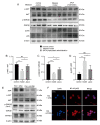
JAC4 inhibits NF-κB (p65) nuclear translocation. (A) Correlation analysis of the expressions of JWA and NF-κB in PD (GSE 6613, n = 50, r = −0.27). (B) The protein expression levels of p-AKT, AKT, p-GSK3β (Ser9), p-GSK3β (Tyr216) and GSK3β. (C) The protein expression levels of NF-κB in the nucleus and cytoplasm. (D) Immunofluorescences image of NF-κB location.
Figure 7
Figure 7
The verification of the mechanism in mice the midbrain and HT-22 cells. (AD) The protein expression levels of p-AKT, AKT, p-GSK3β (Ser9), GSK3β, p-p65 and p65 in the midbrain of mice and their resulting quantized statistics. (E,F) The protein expression levels of JWA, p-AKT, AKT, p-GSK3β (Ser9), GSK3β and MT-ND1. (E) Immunofluorescence images of NF-κB location (F) in HT-22 cells Rot-treated for 48 h after shJWA transfection. The results are shown as the mean ± SEM (n = 3, * p < 0.05, ** p < 0.01, *** p < 0.001, ns p > 0.05).
Figure 8
Figure 8
Schematic diagram of the molecular mechanism of the neuroprotective effect of JAC4 in the Rot-induced PD model.

References

    1. McGregor M.M., Nelson A.B. Circuit Mechanisms of Parkinson’s Disease. Neuron. 2019;101:1042–1056. doi: 10.1016/j.neuron.2019.03.004. - DOI - PubMed
    1. Tolosa E., Garrido A., Scholz S.W., Poewe W. Challenges in the diagnosis of Parkinson’s disease. Lancet Neurol. 2021;20:385–397. doi: 10.1016/S1474-4422(21)00030-2. - DOI - PMC - PubMed
    1. Dorsey E.R., Bloem B.R. The Parkinson Pandemic-A Call to Action. JAMA Neurol. 2018;75:9–10. doi: 10.1001/jamaneurol.2017.3299. - DOI - PubMed
    1. Simon D.K., Tanner C.M., Brundin P. Parkinson Disease Epidemiology, Pathology, Genetics, and Pathophysiology. Clin. Geriatr. Med. 2020;36:1–12. doi: 10.1016/j.cger.2019.08.002. - DOI - PMC - PubMed
    1. Paul K.C., Ritz B. Epidemiology meets toxicogenomics: Mining toxicologic evidence in support of an untargeted analysis of pesticides exposure and Parkinson’s disease. Environ. Int. 2022;170:107613. doi: 10.1016/j.envint.2022.107613. - DOI - PMC - PubMed

LinkOut - more resources