

ABCDEG Stress Echocardiography in Aortic Stenosis
- PMID: 37238211
- PMCID: PMC10217228
- DOI: 10.3390/diagnostics13101727
ABCDEG Stress Echocardiography in Aortic Stenosis
Abstract
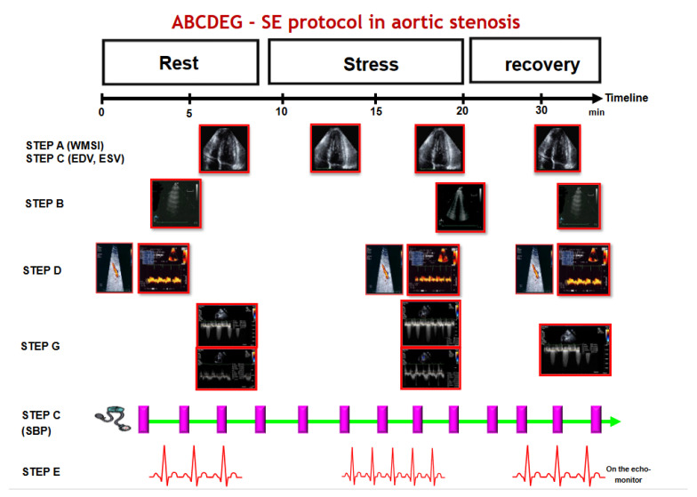
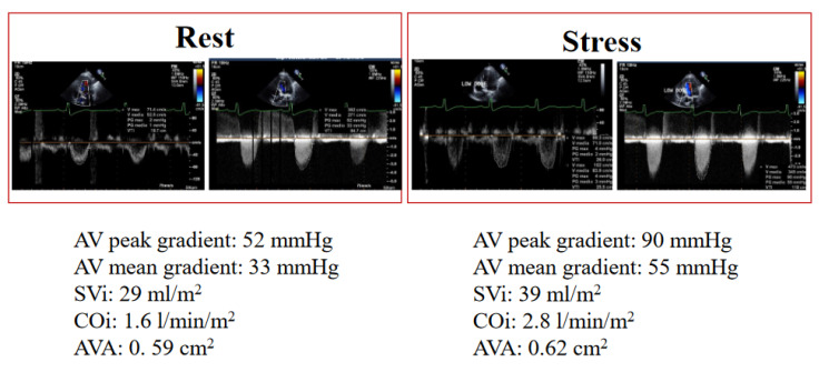
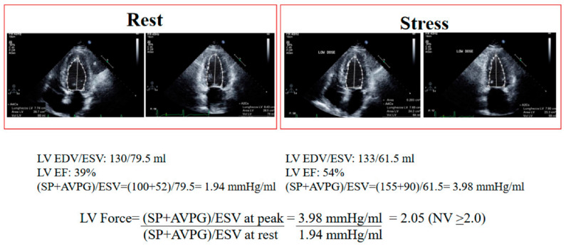
Rest and stress echocardiography (SE) plays a pivotal role in the evaluation of valvular heart disease. The use of SE is recommended in valvular heart disease when there is a mismatch between resting transthoracic echocardiography findings and symptoms. In aortic stenosis (AS), rest echocardiographic analysis is a stepwise approach that begins with the evaluation of aortic valve morphology and proceeds to the measurement of the transvalvular aortic gradient and aortic valve area (AVA) using continuity equations or planimetry. The presence of the following three criteria suggests severe AS: AVA < 1.0 cm2, a peak velocity > 4.0 m/s, or a mean gradient > 40 mmHg. However, in approximately one in three cases, we can observe a discordant AVA < 1 cm2 with a peak velocity < 4.0 m/s or a mean gradient <40 mmHg. This is due to reduced transvalvular flow associated with LV systolic dysfunction (LVEF < 50%) defined as "classical" low-flow low-gradient (LFLG) AS or normal LVEF "paradoxical" LFLG AS. SE has an established role in evaluating LV contractile reserve (CR) patients with reduced LVEF. In classical LFLG AS, LV CR distinguished pseudo-severe AS from truly severe AS. Some observational data suggest that long-term prognosis in asymptomatic severe AS may not be as favorable as previously thought, offering a window of opportunity for intervention prior to the onset of symptoms. Therefore, guidelines recommend evaluating asymptomatic AS with exercise stress in physically active patients, particularly those younger than 70 years, and symptomatic classical LFLG severe AS with low-dose dobutamine SE. A comprehensive SE assessment includes evaluating valve function (gradients), the global systolic function of the LV, and pulmonary congestion. This assessment integrates considerations of blood pressure response, chronotropic reserve, and symptoms. StressEcho 2030 is a prospective, large-scale study that employs a comprehensive protocol (ABCDEG) to analyze the clinical and echocardiographic phenotypes of AS, capturing various vulnerability sources which support stress echo-driven treatment strategies.
Keywords: aortic stenosis; coronary flow reserve; echocardiography; left ventricular contractile reserve; stress echocardiography.
Conflict of interest statement
The authors declare no conflict of interest.
Figures
References
-
- Otto C.M., Nishimura R.A., Bonow R.O., Carabello B.A., Erwin J.P., 3rd, Gentile F., Jneid H., Krieger E.V., Mack M., McLeod C., et al. 2020 ACC/AHA guideline for the management of patients with valvular heart disease: A report of the American College of Cardiology/American Heart Association Joint Committee on Clinical Practice Guidelines. J. Thorac. Cardiovasc. Surg. 2021;143:e183–e353. doi: 10.1016/j.jtcvs.2021.04.002. - DOI - PubMed
-
- Vahanian A., Beyersdorf F., Praz F., Milojevic M., Baldus S., Bauersachs J., Capodanno D., Conradi L., De Bonis M., De Paulis R., et al. ESC/EACTS Scientific Document Group. 2021 ESC/EACTS Guidelines for the management of valvular heart disease. Eur. Heart J. 2022;43:561–632. doi: 10.1093/eurheartj/ehab395. - DOI - PubMed
-
- Knuuti J., Wijns W., Saraste A., Capodanno D., Barbato E., Funck-Brentano C., Prescott E., Storey R.F., Deaton C., Cuisset T., et al. ESC Scientific Document Group 2019 ESC Guidelines for the diagnosis and management of chronic coronary syndromes. Eur. Heart J. 2019;41:407–477. doi: 10.1093/eurheartj/ehz425. - DOI - PubMed
-
- Writing Committee Members. Gulati M., Levy P.D., Mukherjee D., Amsterdam E., Bhatt D.L., Birtcher K.K., Blankstein R., Boyd J., Bullock-Palmer R.P., et al. 2021 AHA/ACC/ASE/ CHEST/SAEM/SCCT/SCMR Guideline for the evaluation and diagnosis of chest pain: A report of the American College of Cardiology/American Heart Association Joint Committee on clinical practice guidelines. J. Cardiovasc. Comput. Tomogr. 2022;16:54122.2013. - PubMed
Publication types
LinkOut - more resources
Full Text Sources
Research Materials
