Case Reports
. 2023 May 19;13(10):1795.
doi: 10.3390/diagnostics13101795.

FIP1L1-PDGFRα-Positive Loeffler Endocarditis-A Distinct Cause of Heart Failure in a Young Male: The Role of Multimodal Diagnostic Tools

Affiliations
Case Reports

FIP1L1-PDGFRα-Positive Loeffler Endocarditis-A Distinct Cause of Heart Failure in a Young Male: The Role of Multimodal Diagnostic Tools

Andreea Varga et al. Diagnostics (Basel). .

Abstract

The presence of the Fip1-Like1-platelet-derived growth factor receptor alpha (FIP1L1-PDGFRα) fusion gene represents a rare cause of hypereosinophilic syndrome (HES), which is associated with organ damage. The aim of this paper is to emphasize the pivotal role of multimodal diagnostic tools in the accurate diagnosis and management of heart failure (HF) associated with HES. We present the case of a young male patient who was admitted with clinical features of congestive HF and laboratory findings of hypereosinophilia (HE). After hematological evaluation, genetic tests, and ruling out reactive causes of HE, a diagnosis of positive FIP1L1-PDGFRα myeloid leukemia was established. Multimodal cardiac imaging identified biventricular thrombi and cardiac impairment, thereby raising suspicion of Loeffler endocarditis (LE) as the cause of HF; this was later confirmed by a pathological examination. Despite hematological improvement under corticosteroid and imatinib therapy, anticoagulant, and patient-oriented HF treatment, there was further clinical progression and subsequent multiple complications (including embolization), which led to patient death. HF is a severe complication that diminishes the demonstrated effectiveness of imatinib in the advanced phases of Loeffler endocarditis. Therefore, the need for an accurate identification of heart failure etiology in the absence of endomyocardial biopsy is particularly important for ensuring effective treatment.

Keywords: FIP1L1–PDGFRA fusion gene; Loeffler endocarditis; cardiac imaging; heart failure; hypereosinophilic syndrome.


Conflict of interest statement

The authors declare no conflict of interest.

Figures

Figure 1
Figure 1
Resting electrocardiogram—sinus rhythm, normal QRS axis, incomplete RBBB, and negative T waves in lateral and inferior leads.
Figure 2
Figure 2
Bidimensional TTE images. (a) The parasternal long axis revealing the presence and extension of the intraventricular mass towards the posterior wall and subvalvular apparatus. (b) Apical four-chamber view (A4C) depicting the hyperechogenic structure obliterating the apex and lateral wall. (c) Apical two-chamber view (A2C) showing the extent of the intraventricular mass to the anterior wall of LV. (d) A4C and color doppler showing moderate-to-severe mitral regurgitation. A—intraventricular mass, AoV—aortic valve, B—interventricular septum, C—lateral wall of the left ventricle, D—inferior wall of the left ventricle, E—anterior wall of the left ventricle, LA—left atrium, LV—left ventricle, LVOT—left ventricular outflow tract, RA—right atrium, and RV—right ventricle.
Figure 3
Figure 3
CMR images revealing left (*) and right (#) intraventricular masses. (a) T1-weighted 4C view. (b) T2-weighted 4C view. (c) T2-weighted fat-suppressed 4C view. (d) 4C-balanced free steady-state precession 4C view. (e,g) Late gadolinium-enhanced SA view. (f) Late gadolinium-enhanced 4C view.
Figure 4
Figure 4
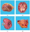
(a) Organized intraventricular thrombi (A and B) are seen in the left (C) and right ventricle (D), respectively. (b,c) Medullar (E) and cortical (F) infarcted areas of the left kidney. (d) Infrarenal aorta (I) with a large saddle thrombus (G) on the aortic bifurcation (H). IVS—interventricular septum and RP—renal pelvis.

References

    1. Valent P., Klion A.D., Horny H.P., Roufosse F., Gotlib J., Weller P.F., Hellmann A., Metzgeroth G., Leiferman K.M., Arock M., et al. Contemporary consensus proposal on criteria and classification of eosinophilic disorders and related syndromes. J. Allergy Clin. Immunol. 2012;130:607–612. doi: 10.1016/j.jaci.2012.02.019. - DOI - PMC - PubMed
    1. Shomali W., Gotlib J. World Health Organization-defined eosinophilic disorders: 2022 update on diagnosis, risk stratification, and management. Am. J. Hematol. 2022;97:129–148. doi: 10.1002/ajh.26352. - DOI - PubMed
    1. Valent P., Degenfeld-Schonburg L., Sadovnik I., Horny H.P., Arock M., Simon H.U., Reiter A., Bochner B.S. Eosinophils and eosinophil-associated disorders: Immunological, clinical, and molecular complexity. Semin. Immunopathol. 2021;43:423–438. doi: 10.1007/s00281-021-00863-y. - DOI - PMC - PubMed
    1. Lupu S., Pop M., Mitre A. Loeffler Endocarditis Causing Heart Failure with Preserved Ejection Fraction (HFpEF): Characteristic Images and Diagnostic Pathway. Diagnostics. 2022;12:2157. doi: 10.3390/diagnostics12092157. - DOI - PMC - PubMed
    1. Requena G., van den Bosch J., Akuthota P., Kovalszki A., Steinfeld J., Kwon N., Van Dyke M.K. Clinical Profile and Treatment in Hypereosinophilic Syndrome Variants: A Pragmatic Review. J. Allergy Clin. Immunol. Pract. 2022;10:2125–2134. doi: 10.1016/j.jaip.2022.03.034. - DOI - PubMed

Publication types