Review
. 2023 May 2;10(5):828.
doi: 10.3390/children10050828.

Fetal Lung Interstitial Tumor (FLIT): Review of The Literature

Affiliations
Review

Fetal Lung Interstitial Tumor (FLIT): Review of The Literature

Silvia Perin et al. Children (Basel). .

Abstract

Fetal lung interstitial tumor (FLIT) is an extremely rare pediatric lung tumor that shares radiological features with congenital pulmonary malformations (cPAM) and other lung neoplasms. A review of the literature, together with the first European case, are herein reported. A systematic and manual search of the literature using the keyword "fetal lung interstitial tumor" was conducted on PUBMED, Scopus, and SCIE (Web of Science). Following the PRISMA guidelines, 12 articles were retrieved which describe a total of 21 cases of FLIT, and a new European case is presented. A prenatal diagnosis was reported in only 3 out of 22 (13%) cases. The mean age at surgery was 31 days of life (1-150); a lobectomy was performed in most of the cases. No complications or recurrence of disease were reported at a mean follow-up of 49 months. FLIT is rarely diagnosed during pregnancy, may present at birth with different levels of respiratory distress, and requires prompt surgical resection. Histology and immunohistochemistry allow for the differentiation of FLIT from cPAM and other lung tumors with poor prognosis, such as pleuropulmonary blastoma, congenital peri-bronchial myofibroblastic tumor, inflammatory myofibroblastic tumor, and congenital or infantile fibrosarcoma.

Keywords: FLIT; congenital pulmonary malformations; lung tumors; respiratory distress.


Conflict of interest statement

The authors declare no conflict of interest.

Figures

Figure 1
Figure 1
PRISMA 2020 flow diagram for new systematic reviews which included searches of databases, registers, and other sources of “fetal lung interstitial tumor” cases.
Figure 2
Figure 2
(A) Gender distribution. (B,C) Tumor location. (D) Age at surgery. (E) Tumor size at resection. (F) Months without recurrence.
Figure 3
Figure 3
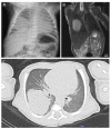
Radiological imaging. (A) X-ray image at 2 days of birth. (B) MRI in T2 phase showing an oval solid mass located at the lower right lobe. (C) TC image confirming the presence of solid mass at the lower right lobe.
Figure 4
Figure 4
FLIT histological findings. (A) FLIT showing bland ovoid interstitial cells with scattered cystic pseudoglandular spaces and thick-walled vessels (hematoxylin eosin ×200). (B) Smooth muscle actin shows only rare myofibroblast-like positive cells (immunohistochemistry, ×200). (C) Almost every cuboidal epithelial cell lining in the pseudoglandular spaces is stained with thyroid transcription factor-1 (immunohistochemistry, ×200). (D) Strong and diffuse vimentin positivity in the interstitial tumor component (immunohistochemistry, ×200).

References

    1. Cass D.L., Olutoye O.O., Cassady C.I., Moise K.J., Johnson A., Papanna R., Lazar D.A., Ayres N.A., Belleza-Bascon B. Prenatal diagnosis and outcome of fetal lung masses. J. Pediatr. Surg. 2011;46:292–298. doi: 10.1016/j.jpedsurg.2010.11.004. - DOI - PubMed
    1. Adams N.C., Victoria T., Oliver E.R., Moldenhauer J.S., Adzick N.S., Colleran G.C. Fetal ultrasound and magnetic resonance imaging: A primer on how to interpret prenatal lung lesions. Pediatr. Radiol. 2020;50:1839–1854. doi: 10.1007/s00247-020-04806-x. - DOI - PubMed
    1. Pogoriler J., Swarr D., Kreiger P., Adzick N.S., Peranteau W. Congenital cystic lung lesions: Redefining the natural distribution of subtypes and assessing the risk of malignancy. Am. J. Surg. Pathol. 2019;43:47. doi: 10.1097/PAS.0000000000000992. - DOI - PMC - PubMed
    1. Waelti S.L., Garel L., Soglio D.D., Rypens F., Messerli M., Dubois J. Neonatal congenital lung tumors—The importance of mid-second-trimester ultrasound as a diagnostic clue. Pediatr. Radiol. 2017;47:1766–1775. doi: 10.1007/s00247-017-3953-3. - DOI - PubMed
    1. Lichtenberger J.P., 3rd, Biko D.M., Carter B.W., Pavio M.A., Huppmann A.R., Chung E.M. Primary Lung Tumors in Children: Radiologic-Pathologic Correlation From the Radiologic Pathology Archives. Radiographics. 2018;38:2151–2172. doi: 10.1148/rg.2018180192. - DOI - PubMed

LinkOut - more resources