. 2023 Apr 23;13(5):726.
doi: 10.3390/biom13050726.

Distinct Effects of Familial Parkinson's Disease-Associated Mutations on α-Synuclein Phase Separation and Amyloid Aggregation

Affiliations

Distinct Effects of Familial Parkinson's Disease-Associated Mutations on α-Synuclein Phase Separation and Amyloid Aggregation

Bingkuan Xu et al. Biomolecules. .

Abstract

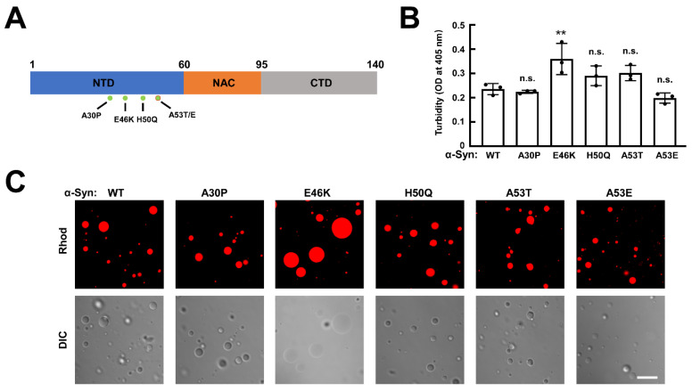
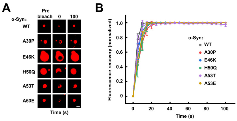
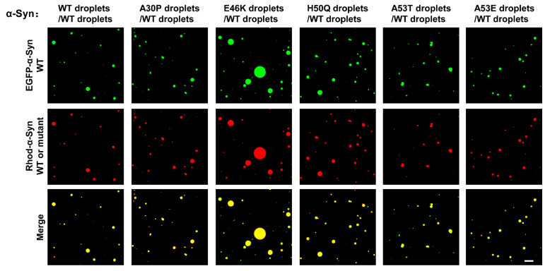
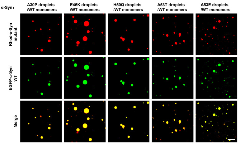
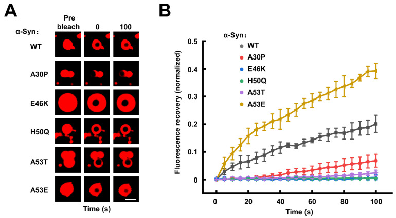
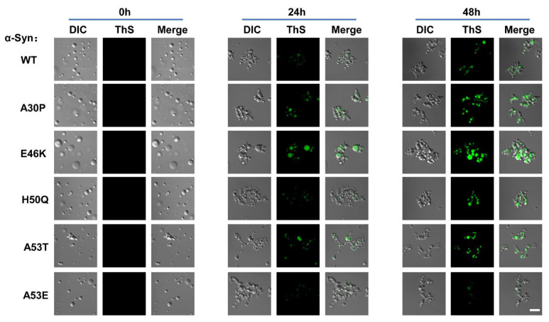
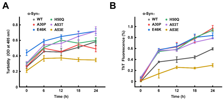
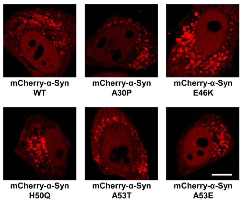
The Lewy bodies and Lewy neurites are key pathological hallmarks of Parkinson's disease (PD). Single-point mutations associated with familial PD cause α-synuclein (α-Syn) aggregation, leading to the formation of Lewy bodies and Lewy neurites. Recent studies suggest α-Syn nucleates through liquid-liquid phase separation (LLPS) to form amyloid aggregates in a condensate pathway. How PD-associated mutations affect α-Syn LLPS and its correlation with amyloid aggregation remains incompletely understood. Here, we examined the effects of five mutations identified in PD, A30P, E46K, H50Q, A53T, and A53E, on the phase separation of α-Syn. All other α-Syn mutants behave LLPS similarly to wild-type (WT) α-Syn, except that the E46K mutation substantially promotes the formation of α-Syn condensates. The mutant α-Syn droplets fuse to WT α-Syn droplets and recruit α-Syn monomers into their droplets. Our studies showed that α-Syn A30P, E46K, H50Q, and A53T mutations accelerated the formation of amyloid aggregates in the condensates. In contrast, the α-Syn A53E mutant retarded the aggregation during the liquid-to-solid phase transition. Finally, we observed that WT and mutant α-Syn formed condensates in the cells, whereas the E46K mutation apparently promoted the formation of condensates. These findings reveal that familial PD-associated mutations have divergent effects on α-Syn LLPS and amyloid aggregation in the phase-separated condensates, providing new insights into the pathogenesis of PD-associated α-Syn mutations.

Keywords: Parkinson’s disease (PD); amyloid aggregation; liquid–liquid phase separation (LLPS); mutation; phase transition; α-synuclein.


Conflict of interest statement

The authors declare no conflict of interest.

Figures

Figure 1
Figure 1
WT and familial PD-associated mutant α-Syn undergo LLPS in vitro. (A) Illustration showing the familial PD-associated mutations. The NTD, NAC, and CTD of α-Syn are indicated in different colors. (B) Turbidity assays showing the formation of phase-separated condensates by WT and mutant α-Syn. Turbidity was evaluated by monitoring the absorbance at 405 nm. Data are presented as mean ± SD (n = 3 independent replicates). p values were calculated using one-way ANOVA with Tukey’s multiple comparisons test. n.s., p > 0.05. **, p < 0.01. (C) Fluorescence and differential interference contrast (DIC) images showing the morphologies of Rhod-labeled WT and mutant α-Syn droplets. The α-Syn concentration is 200 μM. Scale bar, 5 μm. All the experiments were carried out in the presence of 20% PEG-10000.
Figure 2
Figure 2
The familial PD-associated mutations do not affect the fluidity of α-Syn condensates. (A) Representative FRAP images of WT and mutant α-Syn condensates. The fluorescence images of prebleached, bleached (0 s), and bleached after 100 s recovery are shown. Scale bar, 2 μm. (B) The normalized FRAP curves of α-Syn condensates shown in A. Data are presented as mean ± SD (n = 3 independent replicates) and normalized to the maximal prebleach and minimal postbleach fluorescence intensities. The concentration of α-Syn is 200 μM. All the experiments were carried out in the presence of 20% PEG-10000.
Figure 3
Figure 3
The mutant and WT α -Syn droplets can fuse. Representative images showing the fusion of two color-labeled droplets. The EGFP-labeled WT α-Syn droplets and Rhod-labeled WT and mutant α-Syn droplets were gently mixed and visualized through a confocal microscope. Scale bar, 5 μm. The experiments were performed in the presence of 20% PEG-10000.
Figure 4
Figure 4
The familial PD-associated mutant α-Syn droplets recruit α-Syn monomers. Representative images showing the entry of EGFP-labeled α-Syn monomers into Rhod-labeled mutant α-Syn droplets. The total concentrations of α-Syn mutants are 200 μM, and the concentration of EGFP-labeled α-Syn monomers is 10 μM. Scale bar, 5 μm. The experiments were performed in the presence of 20% PEG-10000.
Figure 5
Figure 5
α-Syn undergoes a liquid-to-solid phase transition differently regulated by familial PD-associated mutations. (A) Representative FRAP images of WT and mutant α-Syn condensates after 48 h incubation. The fluorescence images of prebleached, bleached (0 s), and bleached after 100 s recovery are shown. Scale bar, 2 μm. (B) The normalized FRAP curves of α-Syn condensates shown in A. Data are presented as mean ± SD (n = 3 independent replicates) and normalized to the maximal prebleach and minimal postbleach fluorescence intensities. The concentration of α-Syn is 200 μM. All the experiments were carried out in the presence of 20% PEG-10000.
Figure 6
Figure 6
Distinct effects of familial PD-associated mutations on the formation of amyloid aggregates in the condensates. ThS staining images showing the amyloid structure inside the α-Syn condensates. WT or mutant α-Syn condensates were incubated with ThS for 0 h (Left), 24 h (Middle), and 48 h (Right), respectively. Scale bar, 5 μm. The concentration of α-Syn is 200 μM. The experiments were performed in the presence of 20% PEG-10000.
Figure 7
Figure 7
The familial PD-associated mutations have different effects on the amyloid aggregation of α-Syn in the condensation pathway. Kinetic curves of turbidity (A) and ThT fluorescence intensity (B) are shown. Data are presented as mean ± SD (n = 3 independent replicates). In the ThT assays, data are normalized to the maximal and minimal average fluorescence intensities. The concentrations of α-Syn are 200 μM. All the experiments were carried out at 37 °C without shaking in the presence of 20% PEG-10000.
Figure 8
Figure 8
The E46K mutation promotes the formation of α-Syn condensates in cells. Representative confocal images of HeLa cells expressing WT and mutant mCherry-α-Syn, respectively. Scale bar, 5 μm.

Similar articles

Cited by

References

    1. Goedert M., Spillantini M.G., Del Tredici K., Braak H. 100 years of Lewy pathology. Nat. Rev. Neurol. 2013;9:13–24. doi: 10.1038/nrneurol.2012.242. - DOI - PubMed
    1. Spillantini M.G., Schmidt M.L., Lee V.M., Trojanowski J.Q., Jakes R., Goedert M. Alpha-synuclein in Lewy bodies. Nature. 1997;388:839–840. doi: 10.1038/42166. - DOI - PubMed
    1. Karpinar D.P., Balija M.B., Kügler S., Opazo F., Rezaei-Ghaleh N., Wender N., Kim H.Y., Taschenberger G., Falkenburger B.H., Heise H., et al. Pre-fibrillar alpha-synuclein variants with impaired beta-structure increase neurotoxicity in Parkinson’s disease models. EMBO J. 2009;28:3256–3268. doi: 10.1038/emboj.2009.257. - DOI - PMC - PubMed
    1. Volpicelli-Daley L.A., Luk K.C., Patel T.P., Tanik S.A., Riddle D.M., Stieber A., Meaney D.F., Trojanowski J.Q., Lee V.M. Exogenous α-synuclein fibrils induce Lewy body pathology leading to synaptic dysfunction and neuron death. Neuron. 2011;72:57–71. doi: 10.1016/j.neuron.2011.08.033. - DOI - PMC - PubMed
    1. Fusco G., Chen S.W., Williamson P.T.F., Cascella R., Perni M., Jarvis J.A., Cecchi C., Vendruscolo M., Chiti F., Cremades N., et al. Structural basis of membrane disruption and cellular toxicity by α-synuclein oligomers. Science. 2017;358:1440–1443. doi: 10.1126/science.aan6160. - DOI - PubMed

Publication types