

Cellular Response to Bone Morphogenetic Proteins-2 and -7 Covalently Bound to Photocrosslinked Heparin-Diazoresin Multilayer
- PMID: 37238712
- PMCID: PMC10216225
- DOI: 10.3390/biom13050842
Cellular Response to Bone Morphogenetic Proteins-2 and -7 Covalently Bound to Photocrosslinked Heparin-Diazoresin Multilayer
Abstract
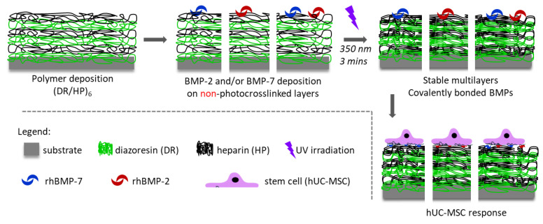
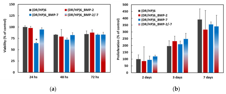
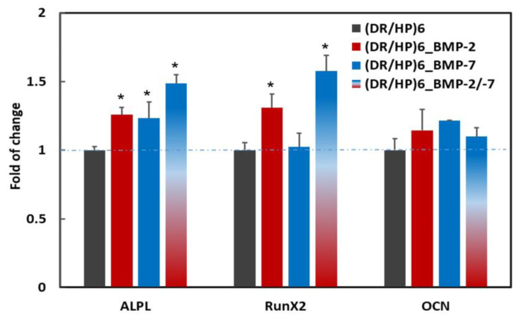
Despite the plethora of research that exists on recombinant human bone morphogenetic protein-2 and -7 (rhBMP-2 and rhBMP-7) and has been clinically approved, there is still a need to gain information that would allow for their more rational use in bone implantology. The clinical application of supra-physiological dosages of these superactive molecules causes many serious adverse effects. At the cellular level, they play a role in osteogenesis and cellular adhesion, migration, and proliferation around the implant. Therefore, in this work, we investigated the role of the covalent binding of rhBMP-2 and rhBMP-7 separately and in combination with ultrathin multilayers composed of heparin and diazoresin in stem cells. In the first step, we optimized the protein deposition conditions via quartz crystal microbalance (QCM). Then, atomic force microscopy (AFM) and enzyme-linked immunosorbent assay (ELISA) were used to analyze protein-substrate interactions. The effect of the protein binding on the initial cell adhesion, migration, and short-term expression of osteogenesis markers was tested. In the presence of both proteins, cell flattening and adhesion became more prominent, resulting in limited motility. However, the early osteogenic marker expression significantly increased compared to the single protein systems. The presence of single proteins resulted in the elongation of cells, which promoted their migration activity.
Keywords: bone morphogenetic protein-2; bone morphogenetic protein-7; cell culture surfaces; diazoresin; heparin.
Conflict of interest statement
The authors declare no conflict of interest.
Figures


Similar articles
-
The Effect of the Topmost Layer and the Type of Bone Morphogenetic Protein-2 Immobilization on the Mesenchymal Stem Cell Response.Int J Mol Sci. 2022 Aug 18;23(16):9287. doi: 10.3390/ijms23169287. Int J Mol Sci. 2022. PMID: 36012551 Free PMC article.
-
Tuning the bioactivity of bone morphogenetic protein-2 with surface immobilization strategies.Acta Biomater. 2018 Oct 15;80:108-120. doi: 10.1016/j.actbio.2018.09.011. Epub 2018 Sep 13. Acta Biomater. 2018. PMID: 30218780
-
Magnesium modification up-regulates the bioactivity of bone morphogenetic protein-2 upon calcium phosphate cement via enhanced BMP receptor recognition and Smad signaling pathway.Colloids Surf B Biointerfaces. 2016 Sep 1;145:140-151. doi: 10.1016/j.colsurfb.2016.04.045. Epub 2016 Apr 26. Colloids Surf B Biointerfaces. 2016. PMID: 27156155
-
N- and E-cadherin mediate early human calvaria osteoblast differentiation promoted by bone morphogenetic protein-2.J Cell Physiol. 2000 Apr;183(1):117-28. doi: 10.1002/(SICI)1097-4652(200004)183:1<117::AID-JCP14>3.0.CO;2-#. J Cell Physiol. 2000. PMID: 10699973
-
Safety profile for the clinical use of bone morphogenetic proteins in the spine.Spine (Phila Pa 1976). 2002 Aug 15;27(16 Suppl 1):S40-8. doi: 10.1097/00007632-200208151-00010. Spine (Phila Pa 1976). 2002. PMID: 12205419 Review.
Cited by
-
Advances in growth factor-containing 3D printed scaffolds in orthopedics.Biomed Eng Online. 2025 Feb 7;24(1):14. doi: 10.1186/s12938-025-01346-z. Biomed Eng Online. 2025. PMID: 39920740 Free PMC article. Review.
References
-
- Brigaud I., Agniel R., Leroy-Dudal J., Kellouche S., Ponche A., Bouceba T., Mihailescu N., Sopronyi M., Viguier E., Ristoscu C., et al. Synergistic effects of BMP-2, BMP-6 or BMP-7 with human plasma fibronectin onto hydroxyapatite coatings: A comparative study. Acta Biomater. 2017;55:481–492. doi: 10.1016/j.actbio.2017.04.013. - DOI - PubMed
Publication types
MeSH terms
Substances
LinkOut - more resources
Full Text Sources
Medical
Miscellaneous
