Review
. 2023 May 18;13(5):853.
doi: 10.3390/biom13050853.

Ribosomal Protein uS5 and Friends: Protein-Protein Interactions Involved in Ribosome Assembly and Beyond

Affiliations
Review

Ribosomal Protein uS5 and Friends: Protein-Protein Interactions Involved in Ribosome Assembly and Beyond

Anne-Marie Landry-Voyer et al. Biomolecules. .

Abstract

Ribosomal proteins are fundamental components of the ribosomes in all living cells. The ribosomal protein uS5 (Rps2) is a stable component of the small ribosomal subunit within all three domains of life. In addition to its interactions with proximal ribosomal proteins and rRNA inside the ribosome, uS5 has a surprisingly complex network of evolutionarily conserved non-ribosome-associated proteins. In this review, we focus on a set of four conserved uS5-associated proteins: the protein arginine methyltransferase 3 (PRMT3), the programmed cell death 2 (PDCD2) and its PDCD2-like (PDCD2L) paralog, and the zinc finger protein, ZNF277. We discuss recent work that presents PDCD2 and homologs as a dedicated uS5 chaperone and PDCD2L as a potential adaptor protein for the nuclear export of pre-40S subunits. Although the functional significance of the PRMT3-uS5 and ZNF277-uS5 interactions remain elusive, we reflect on the potential roles of uS5 arginine methylation by PRMT3 and on data indicating that ZNF277 and PRMT3 compete for uS5 binding. Together, these discussions highlight the complex and conserved regulatory network responsible for monitoring the availability and the folding of uS5 for the formation of 40S ribosomal subunits and/or the role of uS5 in potential extra-ribosomal functions.

Keywords: PDCD2; PDCD2L; PRMT3; ZNF277; dedicated chaperone; ribosome biogenesis; uS5.


Conflict of interest statement

The authors declare no conflict of interest.

Figures

Figure 1
Figure 1
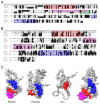
Structural characteristics of the 40S ribosomal protein uS5. (A) Cryo-EM structure of the actively translating 40 ribosomal subunit (PDB entry 5AJ0), left, and rotated 90 degrees, right. uS5 is shown in cyan, while the other 40S ribosomal proteins are colored in grey. The 18S rRNA is shown in orange. (B) Superposition of the tertiary structures of uS5 extracted from the active 40S ribosomal subunit (PDB entry 5AJ0; dark blue, magenta, and orange colors) and predicted by AlphaFold (pale blue). Note that the uS5 structure from the 40S subunit (PDB 5AJ0) represents only amino acids D57 to T278. The double-stranded RNA-binding-like domain and the conserved S5 C-terminal domain are shown in pink and orange, respectively, while the N- and C-terminal extensions are only seen in the AlphaFold model (pale blue). (C) Superposition of the AlphaFold tertiary structures of uS5 from E. coli (lime, P0A7W1), S. cerevisiae (magenta, P25443), and human (cyan, P15880) showing eukaryotic-specific N- and C-terminal extensions. (D) Motifs and functional domains of uS5 are shown. Numbers indicate the amino acid positions of each domain. Alignment and shading were generated using ClustalW and Boxshade software. Sequences are from Homo sapiens (H.s.), Drosophila melanogaster (D.m.), Caenorhabditis elegans (C.e.). Arabidopsis thaliana (A.t.), Saccharomyces cerevisiae (S.c.), and Schizosaccharomyces pombe (S.p.). The FXXXFG and FG motifs are boxed in red.
Figure 2
Figure 2
Sequence and structural analysis of PDCD2 and PDCD2L paralogs. (A) Amino acid sequence alignment of human PDCD2 and PDCD2L. Both proteins harbor N- and C-terminal TYPP domains (highlighted in red and blue, respectively) with conserved GxxP, Cx1-2C, and Q residues highlighted with circles. Whereas PDCD2 contains a MYND zinc finger domain (in magenta with critical cysteine and histidine residues indicated by circles marked above), PDCD2L harbors a leucine-rich NES consensus sequence (green), Φx2-3Φx2-3ΦxΦ, where Φ represents large hydrophobic residues (indicated by green circles marked underneath). (B) S. cerevisiae Tsr4 lacks the MYND zinc finger domain and its C-terminal TYPP domain is degenerated. (C) AlphaFold structures for human PDCD2 (Q16342), human PDCD2L (Q9BRP1), yeast Tsr4 (P25040), and Drosophila Zfrp8 (Q9W1A3). Red: N-terminal TYPP domain; Blue: C-terminal TYPP domain; Magenta: MYND domain.
Figure 3
Figure 3
Predicted model of the human uS5–PDCD2 complex. (A) AlphaFold-Multimer [60] prediction of the human uS5 (cyan)–PDCD2 (orange) complex, left, and rotated 180 degrees, right. (B) AlphaFold-predicted aligned error plot for the uS5 monomer (left) and uS5–PDCD2 complex (right), highlighting residues 20–50 of uS5 confidently predicted to interact with PDCD2 and residues 273–293 that show reduced predicted position error. (C) Surface representation of PDCD2 lipophilicity with ribbon-like structure of uS5 (green), left, and rotated 90 degrees, right. Residues 20–50 of uS5 are colored in magenta. (D) Phe25 and Phe29 residues of human uS5 are predicted to be embedded in hydrophobic core regions of PDCD2. (E) Surface representation of the uS5 (cyan)–PDCD2 (orange) complex, left, and rotated 180 degrees, right. A C-shaped region of PDCD2 (aa 204–239) wraps around the S5 dsRBD of uS5.
Figure 4
Figure 4
Model of how human PDCD2 and PDCD2L contribute to 40S ribosomal subunit biogenesis via interaction with uS5. (1) PDCD2 binds nascent uS5 co-translationally. (2) Interaction between uS5 and PDCD2 takes place in the cytoplasm and the (3) nucleolus. The role of PDCD2 in the nuclear import of uS5 remains to be determined. (4) In the nucleolus, PDCD2 would promote the incorporation of uS5 into the pre-40S ribosomal subunit. (5) PDCD2L binds to pre-40S subunits in the nucleolus via interaction with uS5. (6) The leucine-rich NES of PDCD2L promotes the recruitment of XPO1/CMR1 to the pre-40S particles. (7) Once pre-40S particles are exported to the cytoplasm, (8) PDCD2L and XPO1/CRM1 would dissocociate from 40S precursors.
Figure 5
Figure 5
Predicted model of the human uS5–PRMT3 complex.(A) AlphaFold-Multimer [60] prediction of the human uS5 (grey)–PRMT3 (magenta) complex, left, and rotated 180 degrees, right. The C2H2 zinc finger (aa 48–71) and methyltransferase (aa 217–531) domains are shown in green and red, respectively. (B) The zinc finger domain of PRMT3 does not contact uS5. Shown is the zinc finger (green) of PRMT3 (magenta) with critical cysteine and histidine residues. Human uS5 is shown in grey. (C) The RG-rich region of human uS5 (aa 34–52, shown in dark blue) is located proximally to the catalytic center of the methyltransferase domain (red) of PRMT3. (D) Internal view of the PRMT3 methyltransferase domain with the Glu-338 critical for catalysis shown in yellow and arginine residues 42, 44, and 46 of uS5 shown in dark blue. (E) Western blot analysis using total extracts prepared from three independent clonal lines of HeLa cells deleted for PRMT3 (lanes 2–4 and 6–8) and wild-type (lanes 1 and 5) HeLa cells. Lanes 1–4 were analyzed for total uS5, while lanes 5–8 were analyzed for arginine-methylated uS5 (uS5-mRG).

References

    1. Anger A.M., Armache J.P., Berninghausen O., Habeck M., Subklewe M., Wilson D.N., Beckmann R. Structures of the human and Drosophila 80S ribosome. Nature. 2013;497:80–85. doi: 10.1038/nature12104. - DOI - PubMed
    1. Petrov A.S., Bernier C.R., Hsiao C., Norris A.M., Kovacs N.A., Waterbury C.C., Stepanov V.G., Harvey S.C., Fox G.E., Wartell R.M., et al. Evolution of the ribosome at atomic resolution. Proc. Natl. Acad. Sci. USA. 2014;111:10251–10256. doi: 10.1073/pnas.1407205111. - DOI - PMC - PubMed
    1. Khatter H., Myasnikov A.G., Natchiar S.K., Klaholz B.P. Structure of the human 80S ribosome. Nature. 2015;520:640–645. doi: 10.1038/nature14427. - DOI - PubMed
    1. Melnikov S., Manakongtreecheep K., Soll D. Revising the Structural Diversity of Ribosomal Proteins Across the Three Domains of Life. Mol. Biol. Evol. 2018;35:1588–1598. doi: 10.1093/molbev/msy021. - DOI - PMC - PubMed
    1. Pelletier J., Thomas G., Volarevic S. Ribosome biogenesis in cancer: New players and therapeutic avenues. Nat. Rev. Cancer. 2018;18:51–63. doi: 10.1038/nrc.2017.104. - DOI - PubMed

Publication types

MeSH terms

Grants and funding

LinkOut - more resources