Review
. 2023 Apr 24;11(5):1258.
doi: 10.3390/biomedicines11051258.

Impact of Hyper- and Hypo-Uricemia on Kidney Function

Affiliations
Review

Impact of Hyper- and Hypo-Uricemia on Kidney Function

Junichiro Miake et al. Biomedicines. .

Abstract

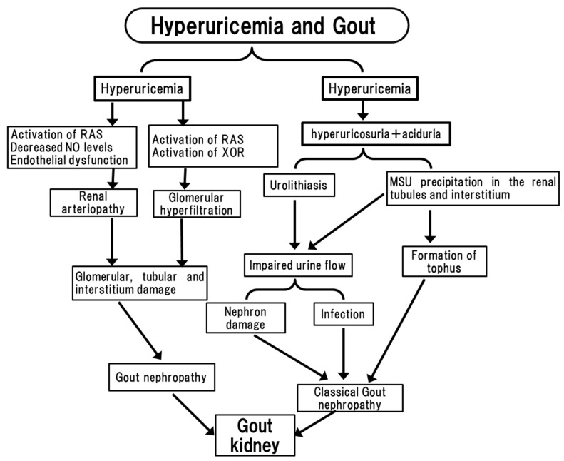
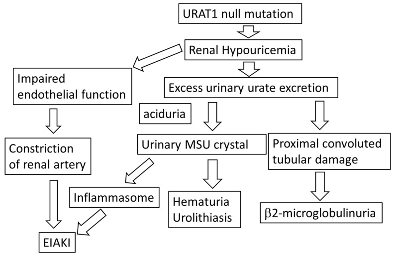
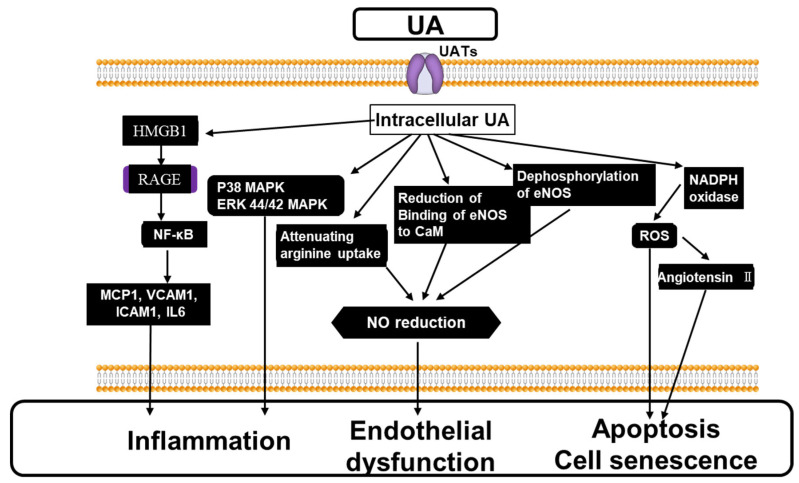
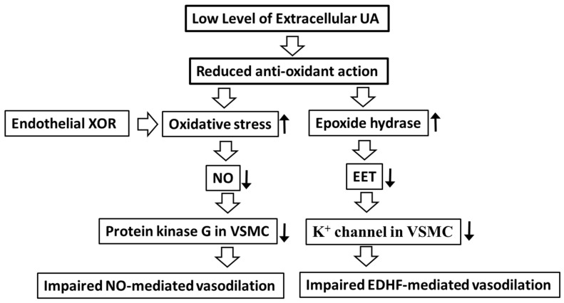
Uric acid (UA) forms monosodium urate (MSU) crystals to exert proinflammatory actions, thus causing gout arthritis, urolithiasis, kidney disease, and cardiovascular disease. UA is also one of the most potent antioxidants that suppresses oxidative stress. Hyper andhypouricemia are caused by genetic mutations or polymorphism. Hyperuricemia increases urinary UA concentration and is frequently associated with urolithiasis, which is augmented by low urinary pH. Renal hypouricemia (RHU) is associated with renal stones by increased level of urinary UA, which correlates with the impaired tubular reabsorption of UA. Hyperuricemia causes gout nephropathy, characterized by renal interstitium and tubular damage because MSU precipitates in the tubules. RHU is also frequently associated with tubular damage with elevated urinary beta2-microglobulin due to increased urinary UA concentration, which is related to impaired tubular UA reabsorption through URAT1. Hyperuricemia could induce renal arteriopathy and reduce renal blood flow, while increasing urinary albumin excretion, which is correlated with plasma xanthine oxidoreductase (XOR) activity. RHU is associated with exercise-induced kidney injury, since low levels of SUA could induce the vasoconstriction of the kidney and the enhanced urinary UA excretion could form intratubular precipitation. A U-shaped association of SUA with organ damage is observed in patients with kidney diseases related to impaired endothelial function. Under hyperuricemia, intracellular UA, MSU crystals, and XOR could reduce NO and activate several proinflammatory signals, impairing endothelial functions. Under hypouricemia, the genetic and pharmacological depletion of UA could impair the NO-dependent and independent endothelial functions, suggesting that RHU and secondary hypouricemia might be a risk factor for the loss of kidney functions. In order to protect kidney functions in hyperuricemic patients, the use of urate lowering agents could be recommended to target SUA below 6 mg/dL. In order to protect the kidney functions in RHU patients, hydration and urinary alkalization may be recommended, and in some cases an XOR inhibitor might be recommended in order to reduce oxidative stress.

Keywords: U-shaped association; endothelial function; hyperuricemia; hypouricemia; kidney disease; tubular disease; uric acid transporters; urolithiasis; xanthine oxidase.


Conflict of interest statement

I.H. got the fund from Fuji pharm. Co., Ltd. (Tokyo, Japan) and Mochida Pharm. Co., Ltd. (Tokyo, Japan).

Figures

Figure 1
Figure 1
Pathophysiology of gout kidney. Hyperuricemia leads to hyperuricosuria-associated aciduria, forming urolithiasis and MSU precipitation. Hyperuricemia also influences several signals to exert renal arteriopathy and glomerular hyperfiltration, causing glomerular, tubular and interstitum damage. RAS: renin angiotensin system, NO: nitric oxide, XOR: xanthine oxidoreductase, MSU: monosodium urate.
Figure 2
Figure 2
Kidney disfunction of RHU. A URAT1 mutation causes RHU. Excess urate excretion causes urinary MSU crystals, resulting in hematuria and urolithiasis. It causes proximal convoluted tubular damage and the elevation of urinary b2-microglobulin. Urinary MSU crystal activates the NLRP3 inflammasome, causing EIAKI. The loss of UA reduces the antioxidant action of the endothelial cells, causing the constriction of the renal artery and EIAKI. URAT1: uric acid transporter 1, MSU: monosodium urate, EIAKI: exercise-induced acute kidney injury.
Figure 3
Figure 3
The mechanism of impaired vasodilation by the elevated extracellular and the resulting intracellular UA. The intracellular UA, through UATs, activates p38 44/42 MAPK, NF-kB, causing inflammation. The intracellular UA reduces NO levels via attenuating arginine uptake, the reduction of binding of eNOS to CaM, and the dephosphorylation of eNOS, leading to impaired endothelial function. The intracellular UA generates superoxide via the activation of NADPH oxidase associated with the production of Angiotensin II, causing apoptosis and cell senescence. See the text for details. CaM: calmodulin; NF-κB: Nuclear Factor-κB, MCP-1: monocyte chemotaxis protein-1, VCAM: vascular cell adhesion molecule, ICAM: intracellular cell adhesion molecule, IL6: interleukin 6, HMGB: High Mobility Group Box, eNOS: endothelial nitric oxide synthesis; NADPH: nicotinamide adenine dinucleotide phosphate; UA: uric acid; UAT: uric acid transporter.
Figure 4
Figure 4
The mechanism of vasodilation promoted by the extracellular UA. The extracellular UA promotes vasodilation via the inhibition of oxidative stress and epoxide hydrolase, which indicates that the low serum UA leads to the impaired vasodilation. See the text for details. EDHF: endothelium-derived highly polarized factors; EET: epoxyeicosatrienoic acids; NO: nitric oxide; UA: uric acid; VCMC: vascular smooth muscle cell.

References

    1. Hisatome I., Li P., Miake J., Taufiq F., Mahati E., Maharani N., Utami S.B., Kuwabara M., Bahrudin U., Ninomiya H. Uric Acid as a Risk Factor for Chronic Kidney Disease and Cardiovascular Disease—Japanese Guideline on the Management of Asymptomatic Hyperuricemia. Circ. J. 2021;85:130–138. doi: 10.1253/circj.CJ-20-0406. - DOI - PubMed
    1. Abou-Elela A. Epidemiology, pathophysiology, and management of uric acid urolithiasis: A narrative review. J. Adv. Res. 2017;8:513–527. doi: 10.1016/j.jare.2017.04.005. - DOI - PMC - PubMed
    1. Huang H., Huang B., Li Y., Huang Y., Li J., Yao H., Jing X., Chen J., Wang J. Uric acid and risk of heart failure: A systematic review and meta-analysis. Eur. J. Heart Fail. 2014;16:15–24. doi: 10.1093/eurjhf/hft132. - DOI - PubMed
    1. Kuwabara M., Niwa K., Nishihara S., Nishi Y., Takahashi O., Kario K., Yamamoto K., Yamashita T., Hisatome I. Hyperuricemia is an independent competing risk factor for atrial fibrillation. Int. J. Cardiol. 2017;231:137–142. doi: 10.1016/j.ijcard.2016.11.268. - DOI - PubMed
    1. Li M., Hu X., Fan Y., Li K., Zhang X., Hou W., Tang Z. Hyperuricemia and the risk for coronary heart disease morbidity and mortality a systematic review and dose-response meta-analysis. Sci. Rep. 2016;6:19520. doi: 10.1038/srep19520. - DOI - PMC - PubMed

LinkOut - more resources